The heart is a muscular organ in most animals. This organ pumps blood through the blood vessels of the circulatory system. The pumped blood carries oxygen and nutrients to the body, while carrying metabolic waste such as carbon dioxide to the lungs. In humans, the heart is approximately the size of a closed fist and is located between the lungs, in the middle compartment of the chest, called the mediastinum.

The circulatory system is a system of organs that includes the heart, blood vessels, and blood which is circulated throughout the entire body of a human or other vertebrate. It includes the cardiovascular system, or vascular system, that consists of the heart and blood vessels. The circulatory system has two divisions, a systemic circulation or circuit, and a pulmonary circulation or circuit. Some sources use the terms cardiovascular system and vascular system interchangeably with the circulatory system.

Coronary circulation is the circulation of blood in the arteries and veins that supply the heart muscle (myocardium). Coronary arteries supply oxygenated blood to the heart muscle. Cardiac veins then drain away the blood after it has been deoxygenated. Because the rest of the body, and most especially the brain, needs a steady supply of oxygenated blood that is free of all but the slightest interruptions, the heart is required to function continuously. Therefore its circulation is of major importance not only to its own tissues but to the entire body and even the level of consciousness of the brain from moment to moment. Interruptions of coronary circulation quickly cause heart attacks, in which the heart muscle is damaged by oxygen starvation. Such interruptions are usually caused by coronary ischemia linked to coronary artery disease, and sometimes to embolism from other causes like obstruction in blood flow through vessels.

A ventricle is one of two large chambers located toward the bottom of the heart that collect and expel blood towards the peripheral beds within the body and lungs. The blood pumped by a ventricle is supplied by an atrium, an adjacent chamber in the upper heart that is smaller than a ventricle. Interventricular means between the ventricles, while intraventricular means within one ventricle.

A pulmonary artery is an artery in the pulmonary circulation that carries deoxygenated blood from the right side of the heart to the lungs. The largest pulmonary artery is the main pulmonary artery or pulmonary trunk from the heart, and the smallest ones are the arterioles, which lead to the capillaries that surround the pulmonary alveoli.

The pulmonary circulation is a division of the circulatory system in all vertebrates. The circuit begins with deoxygenated blood returned from the body to the right atrium of the heart where it is pumped out from the right ventricle to the lungs. In the lungs the blood is oxygenated and returned to the left atrium to complete the circuit.

The jugular veins are veins that take blood from the head back to the heart via the superior vena cava. The internal jugular vein descends next to the internal carotid artery and continues posteriorly to the sternocleidomastoid muscle.
The atrium is one of the two upper chambers in the heart that receives blood from the circulatory system. The blood in the atria is pumped into the heart ventricles through the atrioventricular mitral and tricuspid heart valves.

In the brain, the interventricular foramina are channels that connect the paired lateral ventricles with the third ventricle at the midline of the brain. As channels, they allow cerebrospinal fluid (CSF) produced in the lateral ventricles to reach the third ventricle and then the rest of the brain's ventricular system. The walls of the interventricular foramina also contain choroid plexus, a specialized CSF-producing structure, that is continuous with that of the lateral and third ventricles above and below it.

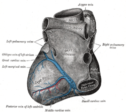
The coronary sinus is the largest vein of the heart. It drains over half of the deoxygenated blood from the heart muscle into the right atrium. It begins on the backside of the heart, in between the left atrium, and left ventricle; it begins at the junction of the great cardiac vein, and oblique vein of the left atrium. It receives multiple tributaries. It passes across the backside of the heart along a groove between left atrium and left ventricle, then drains into the right atrium at the orifice of the coronary sinus.

The great cardiac vein is a vein of the heart. It begins at the apex of the heart and ascends along the anterior interventricular sulcus before joining the oblique vein of the left atrium to form the coronary sinus upon the posterior surface of the heart.
The small cardiac vein, also known as the right coronary vein, is a coronary vein that drains parts of the right atrium and right ventricle of the heart. Despite its size, it is one of the major drainage vessels for the heart.
The middle cardiac vein commences at the apex of the heart. It passes posteriorly along the inferior interventricular sulcus to end at the coronary sinus near the sinus' termination.

The sinus venosus is a large quadrangular cavity which precedes the atrium on the venous side of the chordate heart.

The anterior interventricular sulcus is one of two grooves separating the ventricles of the heart. They can also be known as paraconal interventricular groove or subsinosal interventricular groove respectively. It is situated on the sternocostal surface of the heart, close to the left margin of the heart. It extends between the coronary sulcus, and the apex of the heart; upon reaching the diaphragmatic surface of the heart, it ends at the notch of cardiac apex. It contains the anterior interventricular branch of the left coronary artery, and great cardiac vein.

The internal cerebral veins are two veins included in the group of deep cerebral veins that drain the deep parts of the hemispheres; each internal cerebral vein is formed near the interventricular foramina by the union of the superior thalamostriate vein and the superior choroid vein.
The left marginal vein is a vein of the heart which courses near or over the left margin of the heart. It drains venous blood from much of the myocardium of the left ventricle. It usually empties into the great cardiac vein.
The anterior cardiac veins are a variable number of small veins which drain blood from the anterior portion of the right ventricle into the right atrium.
The smallest cardiac veins are small, valveless veins in the walls of all four heart chambers that drain venous blood from the myocardium directly into any of the heart chambers.

Heart development, also known as cardiogenesis, refers to the prenatal development of the heart. This begins with the formation of two endocardial tubes which merge to form the tubular heart, also called the primitive heart tube. The heart is the first functional organ in vertebrate embryos.