The intercostal muscles comprise many different groups of muscles that run between the ribs, and help form and move the chest wall. The intercostal muscles are mainly involved in the mechanical aspect of breathing by helping expand and shrink the size of the chest cavity.

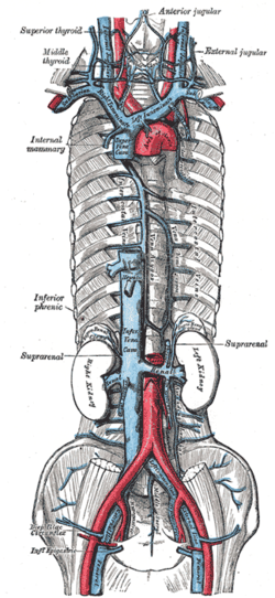
The azygos vein is a vein running up the right side of the thoracic vertebral column draining itself towards the superior vena cava. It connects the systems of superior vena cava and inferior vena cava and can provide an alternative path for blood to the right atrium when either of the venae cavae is blocked.

The internal thoracic artery (ITA), also known as the internal mammary artery, is an artery that supplies the anterior chest wall and the breasts. It is a paired artery, with one running along each side of the sternum, to continue after its bifurcation as the superior epigastric and musculophrenic arteries.

In human anatomy, the internal thoracic vein is the vein that drains the chest wall and breasts.

The superior sagittal sinus, within the human head, is an unpaired area along the attached margin of the falx cerebri. It allows blood to drain from the lateral aspects of anterior cerebral hemispheres to the confluence of sinuses. Cerebrospinal fluid drains through arachnoid granulations into the superior sagittal sinus and is returned to venous circulation.

The hemiazygos vein is a vein running superiorly in the lower thoracic region, just to the left side of the vertebral column.
The accessory hemiazygos vein, also called the superior hemiazygous vein, is a vein on the left side of the vertebral column that generally drains the fourth through eighth intercostal spaces on the left side of the body.

The intercostal space (ICS) is the anatomic space between two ribs. Since there are 12 ribs on each side, there are 11 intercostal spaces, each numbered for the rib superior to it.
The subcostal arteries, so named because they lie below the last ribs, constitute the lowest pair of branches derived from the thoracic aorta, and are in series with the intercostal arteries.

The middle colic vein drains the transverse colon. It is a tributary of the superior mesenteric vein, and follows the path of its corresponding artery, the middle colic artery. As the superior mesenteric vein drains to the hepatic portal vein, the middle colic vein is considered part of the hepatic portal system. This vein also carries nutrients absorbed from the large intestine to the liver.
The right colic vein drains the ascending colon, and is a tributary of the superior mesenteric vein. It travels with its corresponding artery, the right colic artery.

The carotid triangle is a portion of the anterior triangle of the neck.
The bronchial veins are small vessels that return blood from the larger bronchi and structures at the roots of the lungs. The right side drains into the azygos vein, while the left side drains into the left superior intercostal vein or the accessory hemiazygos vein. Bronchial veins are thereby part of the bronchial circulation, carrying waste products away from the cells that constitute the lungs.
The superior intercostal veins are two veins that drain the 2nd, 3rd, and 4th intercostal spaces, one vein for each side of the body.
The posterior intercostal veins are veins that drain the intercostal spaces posteriorly. They run with their corresponding posterior intercostal artery on the underside of the rib, the vein superior to the artery. Each vein also gives off a dorsal branch that drains blood from the muscles of the back.
The intercostal arteries are a group of arteries passing within an intercostal space. There are 9 anterior and 11 posterior intercostal arteries on each side of the body. The anterior intercostal arteries are branches of the internal thoracic artery and its terminal branch - the musculophrenic artery. The posterior intercostal arteries are branches of the supreme intercostal artery and thoracic aorta.

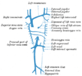
The lumbar veins are four pairs of veins running along the inside of the posterior abdominal wall, and drain venous blood from parts of the abdominal wall. Each lumbar vein accompanies a single lumbar artery. The lower two pairs of lumbar veins all drain directly into the inferior vena cava, whereas the fate of the upper two pairs is more variable.

The intervertebral veins accompany the spinal nerves through the intervertebral foramina to drain the internal vertebral venous plexuses into the external vertebral venous plexuses. They drain into vertebral vein, intercostal veins, lumbar veins, and lateral sacral veins. Upper posterior intercostal veins may additionally drain via brachiocephalic vens. They may drain to ascending lumbar veins. They may drain into the inferior vena cava directly, reaching it by winding around the surface of the vertebral body.

The uterine plexuses lie along the sides and superior angles of the uterus between the two layers of the broad ligament, and communicate with the ovarian and vaginal plexuses.
The intercostal veins are a group of veins which drain the area between the ribs ("costae"), called the intercostal space.