 |
| Part of a series of lists about |
| Human anatomy |
|---|
Depression, in an anatomical term of motion for movement in an inferior direction. It is the opposite of elevation.
| |
| Part of a series of lists about |
| Human anatomy |
|---|
Depression, in an anatomical term of motion for movement in an inferior direction. It is the opposite of elevation.

The trapezius is a large paired trapezoid-shaped surface muscle that extends longitudinally from the occipital bone to the lower thoracic vertebrae of the spine and laterally to the spine of the scapula. It moves the scapula and supports the arm.

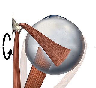
The superior oblique muscle or obliquus oculi superior is a fusiform muscle originating in the upper, medial side of the orbit which abducts, depresses and internally rotates the eye. It is the only extraocular muscle innervated by the trochlear nerve.

The omohyoid muscle is a muscle in the neck. It is one of the infrahyoid muscles. It consists of two bellies separated by an intermediate tendon. Its inferior belly is attached to the scapula; its superior belly is attached to the hyoid bone. Its intermediate tendon is anchored to the clavicle and first rib by a fascial sling. The omohyoid is innervated by the ansa cervicalis of the cervical plexus. It acts to depress the hyoid bone.

The digastric muscle is a bilaterally paired suprahyoid muscle located under the jaw. Its posterior belly is attached to the mastoid notch of temporal bone, and its anterior belly is attached to the digastric fossa of mandible; the two bellies are united by an intermediate tendon which is held in a loop that attaches to the hyoid bone. The anterior belly is innervated via the mandibular nerve, and the posterior belly is innervated via the facial nerve. It may act to depress the mandible or elevate the hyoid bone.
The inferior rectus muscle is a muscle in the orbit near the eye. It is one of the four recti muscles in the group of extraocular muscles. It originates from the common tendinous ring, and inserts into the anteroinferior surface of the eye. It depresses the eye (downwards).

The lips are a horizontal pair of soft appendages attached to the jaws and are the most visible part of the mouth of many animals, including humans. Vertebrate lips are soft, movable and serve to facilitate the ingestion of food and the articulation of sound and speech. Human lips are also a somatosensory organ, and can be an erogenous zone when used in kissing and other acts of intimacy.

The lateral pterygoid muscle (or external pterygoid muscle) is a muscle of mastication. It has two heads. It lies superior to the medial pterygoid muscle. It is supplied by pterygoid branches of the maxillary artery, and the lateral pterygoid nerve (from the mandibular nerve, CN V3). It depresses and protrudes the mandible. When each muscle works independently, they can move the mandible side to side.

The mylohyoid muscle or diaphragma oris is a paired muscle of the neck. It runs from the mandible to the hyoid bone, forming the floor of the oral cavity of the mouth. It is named after its two attachments near the molar teeth. It forms the floor of the submental triangle. It elevates the hyoid bone and the tongue, important during swallowing and speaking.

The depressor supercilii is an eye muscle of the human body. The nature of this muscle is in some dispute. Few printed anatomies include it and many authorities consider it to be part of the orbicularis oculi muscle.

The sternohyoid muscle is a bilaterally paired, long, thin, narrow strap muscle of the anterior neck. It is one of the infrahyoid muscles. It is innervated by the ansa cervicalis. It acts to depress the hyoid bone.

The sternothyroid muscle is an infrahyoid muscle of the neck. It acts to depress the hyoid bone.

The thyrohyoid muscle is a small skeletal muscle of the neck. Above, it attaches onto the greater cornu of the hyoid bone; below, it attaches onto the oblique line of the thyroid cartilage. It is innervated by fibres derived from the cervical spinal nerve 1 that run with the hypoglossal nerve to reach this muscle. The thyrohyoid muscle depresses the hyoid bone and elevates the larynx during swallowing. By controlling the position and shape of the larynx, it aids in making sound.

The internal intercostal muscles are a group of skeletal muscles located between the ribs. They are eleven in number on either side. They commence anteriorly at the sternum, in the intercostal spaces between the cartilages of the true ribs, and at the anterior extremities of the cartilages of the false ribs, and extend backward as far as the angles of the ribs, hence they are continued to the vertebral column by thin aponeuroses, the posterior intercostal membranes. They pull the sternum and ribs upward and inward.

The subclavius is a small triangular muscle, placed between the clavicle and the first rib. Along with the pectoralis major and pectoralis minor muscles, the subclavius muscle makes up the anterior axioappendicular muscles, also known as anterior wall of the axilla.

The hyoglossus is a thin and quadrilateral extrinsic muscle of the tongue. It originates from the hyoid bone; it inserts onto the side of the tongue. It is innervated by the hypoglossal nerve. It acts to depress and retract the tongue.

The mental foramen is one of two foramina (openings) located on the anterior surface of the mandible. It is part of the mandibular canal. It transmits the terminal branches of the inferior alveolar nerve and the mental vessels.

The marginal mandibular branch of the facial nerve arises from the facial nerve in the parotid gland at the parotid plexus. It passes anterior-ward deep to the platysma and depressor anguli oris muscles. It provides motor innervation to muscles of the lower lip and chin: the depressor labii inferioris muscle, depressor anguli oris muscle, and mentalis muscle. It communicates with the mental branch of the inferior alveolar nerve.

The submental artery is the largest branch of the facial artery in the neck. It first runs forward under the mouth, then turns upward upon reaching the chin.

The buccal space is a fascial space of the head and neck. It is a potential space in the cheek, and is paired on each side. The buccal space is superficial to the buccinator muscle and deep to the platysma muscle and the skin. The buccal space is part of the subcutaneous space, which is continuous from head to toe.

The following outline is provided as an overview of and topical guide to human anatomy: