Depressor labii | |
---|---|
![]() Muscles of the head, face, and neck. | |
Details | |
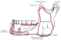
Origin | Oblique line of the mandible, between the symphysis and the mental foramen |
Insertion | Integument of the lower lip, orbicularis oris fibers, its fellow of the opposite side |
Nerve | Marginal mandibular branch of the facial nerve |
Actions | Depression of the lower lip |
Antagonist | Orbicularis oris muscle |
Identifiers | |
Latin | musculus depressor labii inferioris |
TA98 | A04.1.03.033 |
TA2 | 2083 |
FMA | 46816 |
Anatomical terms of muscle |
The depressor labii inferioris (or quadratus labii inferioris) is a facial muscle. It helps to lower the bottom lip.
The depressor labii inferioris muscle arises from the lateral surface of the mandible. [1] This is below the mental foramen, and the origin may be around 3 cm wide. [1] It inserts on the skin of the lower lip, blending in with the orbicularis oris muscle around 2 cm wide. [1] At its origin, depressor labii is continuous with the fibers of the platysma muscle. Some yellow fat is intermingled with the fibers.
The depressor labii inferioris muscle is supplied by the marginal mandibular branch of the facial nerve. [1] [2]
The depressor labii inferioris muscle helps to depress and everts the lower lip. [1] It is the most important of the muscles of the lower lip for this function. [1] It is an antagonist of the orbicularis oris muscle. [1] It is needed to expose the mandibular (lower) teeth during smiling. [1]
The depressor labii inferioris muscle may be resected (cut and removed) using surgery to correct an asymmetry of the lower lip when smiling. [1] This asymmetry can be caused by paralysis of the marginal mandibular branch of the facial nerve on one side, so the healthy side may be cut to create symmetry. [1] Local anaesthesia may be used, such as by blocking the mental nerve. [1] This operation tends to be successful. [1]
The depressor labii inferioris muscle has also (mainly historically) been called the quadratus labii inferioris muscle. [1]
The facial nerve, also known as the seventh cranial nerve, cranial nerve VII, or simply CN VII, is a cranial nerve that emerges from the pons of the brainstem, controls the muscles of facial expression, and functions in the conveyance of taste sensations from the anterior two-thirds of the tongue. The nerve typically travels from the pons through the facial canal in the temporal bone and exits the skull at the stylomastoid foramen. It arises from the brainstem from an area posterior to the cranial nerve VI and anterior to cranial nerve VIII.
The buccinator is a thin quadrilateral muscle occupying the interval between the maxilla and the mandible at the side of the face. It forms the anterior part of the cheek or the lateral wall of the oral cavity.
In human anatomy, the orbicularis oris muscle is a complex of muscles in the lips that encircles the mouth. It is not a true sphincter, as was once thought, as it is actually composed of four independent quadrants that interlace and give only an appearance of circularity.
The lips are a horizontal pair of soft appendages attached to the jaws and are the most visible part of the mouth of many animals, including humans. Vertebrate lips are soft, movable and serve to facilitate the ingestion of food and the articulation of sound and speech. Human lips are also a somatosensory organ, and can be an erogenous zone when used in kissing and other acts of intimacy.
The platysma muscle is a superficial muscle of the human neck that overlaps the sternocleidomastoid. It covers the anterior surface of the neck superficially. When it contracts, it produces a slight wrinkling of the neck, and a "bowstring" effect on either side of the neck.
The zygomaticus major muscle is a muscle of the face. It arises from either zygomatic arch (cheekbone); it inserts at the corner of the mouth. It is innervated by branches of the facial nerve.
The levator labii superioris is a muscle of the human body used in facial expression. It is a broad sheet, the origin of which extends from the side of the nose to the zygomatic bone.
The mentalis muscle is a paired central muscle of the lower lip, situated at the tip of the chin. It originates from the mentum of the mandible, and inserts into the soft tissue of the chin. It is sometimes referred to as the "pouting muscle" due to it raising the lower lip and causing chin wrinkles.
The depressor anguli oris muscle is a facial muscle. It originates from the mandible and inserts into the angle of the mouth. It is associated with frowning, as it depresses the corner of the mouth.
The facial artery is a branch of the external carotid artery that supplies structures of the superficial face.
In facial anatomy, the modiolus is a dense, compact, mobile, fibromuscular tissue mass of facial muscles formed by the interlacing of a number of muscles just lateral to the angle of the mouth opposite the second upper premolar tooth.
The mental foramen is one of two foramina (openings) located on the anterior surface of the mandible. It is part of the mandibular canal. It transmits the terminal branches of the inferior alveolar nerve and the mental vessels.
The mental nerve is a sensory nerve of the face. It is a branch of the posterior trunk of the inferior alveolar nerve, itself a branch of the mandibular nerve (CN V3), itself a branch of the trigeminal nerve (CN V). It provides sensation to the front of the chin and the lower lip, as well as the gums of the anterior mandibular (lower) teeth. It can be blocked with local anaesthesia for procedures on the chin, lower lip, and mucous membrane of the inner cheek. Problems with the nerve cause chin numbness.
The marginal mandibular branch of the facial nerve arises from the facial nerve in the parotid gland at the parotid plexus. It passes anterior-ward deep to the platysma and depressor anguli oris muscles. It provides motor innervation to muscles of the lower lip and chin: the depressor labii inferioris muscle, depressor anguli oris muscle, and mentalis muscle. It communicates with the mental branch of the inferior alveolar nerve.
The buccal branches of the facial nerve, are of larger size than the rest of the branches, pass horizontally forward to be distributed below the orbit and around the mouth.
The submental artery is the largest branch of the facial artery in the neck. It first runs forward under the mouth, then turns upward upon reaching the chin.
The facial muscles are a group of striated skeletal muscles supplied by the facial nerve that, among other things, control facial expression. These muscles are also called mimetic muscles. They are only found in mammals, although they derive from neural crest cells found in all vertebrates. They are the only muscles that attach to the dermis.
Lip reconstruction may be required after trauma or surgical excision. The lips are considered the beginning of the oral cavity and are the most common site of oral cancer. Any reconstruction of the lips must include both functional and cosmetic considerations. The lips are necessary for speech, facial expression, and eating. Because of their prominent location on the face, even small abnormalities can be apparent.
Jaw reduction or mandible angle reduction is a type of surgery to narrow the lower one-third of the face—particularly the contribution from the mandible and its muscular attachments. There are several techniques for treatment—including surgical and non-surgical methods. A square lower jaw can be considered a masculine trait, especially in Asian countries. As a result, whereas square lower jaws are often considered a positive trait in men, a wide mandible can be perceived as discordant or masculine on women, or sometimes in certain men, particularly when there is asymmetry.
In jawed vertebrates, the mandible, lower jaw, or jawbone is a bone that makes up the lower – and typically more mobile – component of the mouth.
This article incorporates text in the public domain from page 383 of the 20th edition of Gray's Anatomy (1918)