Styloglossus

Last updated
Styloglossus
Styloglossus.png
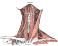
Extrinsic muscles of the tongue. Left side. (Styloglossus XII visible at center top.)
Gray1020.png
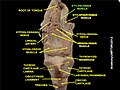
Coronal section of tongue, showing intrinsic muscles. (Styloglossus labeled at center left.)
Details
Origin Styloid process of temporal bone
Insertion Tip and sides of tongue
Artery Sublingual branch of the lingual artery
Nerve Hypoglossal nerve (CN XII)
Actions Retraction and elevation of tongue
Identifiers
Latin musculus styloglossus
TA98 A05.1.04.105
TA2 2121
FMA 46692
Anatomical terms of muscle

The styloglossus muscle is a bilaterally paired muscle of the tongue. It originates at the styloid process of the temporal bone. It inserts onto the side of the tongue. It acts to elevate and retract the tongue. It is innervated by the hypoglossal nerve (cranial nerve XII). [1]

Contents

Anatomy

The styloglossus muscle is the shortest and smallest of the three styloid muscles.[ citation needed ]

Origin

It arises from (the anterior and lateral surfaces of) the styloid process of the temporal bone near its apex, and from the stylomandibular ligament.[ citation needed ]

Course and relations

It passes anterioinferiorly from its origin to its insertion between the internal carotid artery and the external carotid artery,[ citation needed ] and between the superior pharyngeal constrictor muscle and the middle pharyngeal constrictor muscle. [1]

Insertion

It divides upon the side of the tongue near the dorsal surface of the tongue, blending with the fibers of the longitudinalis inferior muscle anterior to the hyoglossus muscle.[ citation needed ]

Innervation

The styloglossus is innervated by the hypoglossal nerve (CN XII) (like all muscles of the tongue except palatoglossus which is innervated by the pharyngeal plexus of vagus nerve (CN X)).[ citation needed ]

Function

The styloglossus draws up the sides of the tongue to create a trough for swallowing. Acting bilaterally (both styloglossus muscles contracting simultaneously) they also aid in retracting the tongue.[ citation needed ]

Additional images

Related Research Articles

<span class="mw-page-title-main">Tongue</span> Muscular organ in the mouth of most vertebrates

The tongue is a muscular organ in the mouth of a typical tetrapod. It manipulates food for chewing and swallowing as part of the digestive process, and is the primary organ of taste. The tongue's upper surface (dorsum) is covered by taste buds housed in numerous lingual papillae. It is sensitive and kept moist by saliva and is richly supplied with nerves and blood vessels. The tongue also serves as a natural means of cleaning the teeth. A major function of the tongue is the enabling of speech in humans and vocalization in other animals.

Articles related to anatomy include:

<span class="mw-page-title-main">Glossopharyngeal nerve</span> Cranial nerve IX, for the tongue and pharynx

The glossopharyngeal nerve, also known as the ninth cranial nerve, cranial nerve IX, or simply CN IX, is a cranial nerve that exits the brainstem from the sides of the upper medulla, just anterior to the vagus nerve. Being a mixed nerve (sensorimotor), it carries afferent sensory and efferent motor information. The motor division of the glossopharyngeal nerve is derived from the basal plate of the embryonic medulla oblongata, whereas the sensory division originates from the cranial neural crest.

<span class="mw-page-title-main">Hypoglossal nerve</span> Cranial nerve XII, for the tongue

The hypoglossal nerve, also known as the twelfth cranial nerve, cranial nerve XII, or simply CN XII, is a cranial nerve that innervates all the extrinsic and intrinsic muscles of the tongue except for the palatoglossus, which is innervated by the vagus nerve. CN XII is a nerve with a sole motor function. The nerve arises from the hypoglossal nucleus in the medulla as a number of small rootlets, pass through the hypoglossal canal and down through the neck, and eventually passes up again over the tongue muscles it supplies into the tongue.

<span class="mw-page-title-main">Internal carotid artery</span> Artery of the human brain

The internal carotid artery is an artery in the neck which supplies the anterior circulation of the brain.

<span class="mw-page-title-main">Digastric muscle</span> Small muscle located under the jaw in mammals

The digastric muscle is a bilaterally paired suprahyoid muscle located under the jaw. Its posterior belly is attached to the mastoid notch of temporal bone, and its anterior belly is attached to the digastric fossa of mandible; the two bellies are united by an intermediate tendon which is held in a loop that attaches to the hyoid bone. The anterior belly is innervated via the mandibular nerve, and the posterior belly is innervated via the facial nerve. It may act to depress the mandible or elevate the hyoid bone.

<span class="mw-page-title-main">Stylohyoid muscle</span> Muscle

The stylohyoid muscle is one of the suprahyoid muscles. Its originates from the styloid process of the temporal bone; it inserts onto hyoid bone. It is innervated by a branch of the facial nerve. It acts draw the hyoid bone upwards and backwards.

<span class="mw-page-title-main">Ansa cervicalis</span> From cervical plexus

The ansa cervicalis is a loop formed by muscular branches of the cervical plexus formed by branches of cervical spinal nerves C1-C3. The ansa cervicalis has two roots - a superior root and an inferior root - that unite distally, forming a loop. It is situated within the carotid sheath.

<span class="mw-page-title-main">Hyoglossus</span> Muscle

The hyoglossus is a thin and quadrilateral extrinsic muscle of the tongue. It originates from the hyoid bone; it inserts onto the side of the tongue. It is innervated by the hypoglossal nerve. It acts to depress and retract the tongue.

<span class="mw-page-title-main">Palatoglossus muscle</span> Muscle of the soft palate

The palatoglossal muscle is a muscle of the soft palate and an extrinsic muscle of the tongue. Its surface is covered by oral mucosa and forms the visible palatoglossal arch.

<span class="mw-page-title-main">Levator veli palatini</span> Muscle of the soft palate

The levator veli palatini is a muscle of the soft palate and pharynx. It is innervated by the vagus nerve via its pharyngeal plexus. During swallowing, it contracts, elevating the soft palate to help prevent food from entering the nasopharynx.

<span class="mw-page-title-main">Middle pharyngeal constrictor muscle</span> Muscle in the neck

The middle pharyngeal constrictor is a fan-shaped muscle located in the neck. It is one of three pharyngeal constrictor muscles. It is smaller than the inferior pharyngeal constrictor muscle.

<span class="mw-page-title-main">Superior pharyngeal constrictor muscle</span> Muscle

The superior pharyngeal constrictor muscle is a quadrilateral muscle of the pharynx. It is the uppermost and thinnest of the three pharyngeal constrictors.

<span class="mw-page-title-main">Stylopharyngeus muscle</span> Muscle

The stylopharyngeus muscle is a muscle in the head. It originates from the temporal styloid process. Some of its fibres insert onto the thyroid cartilage, while others end by intermingling with proximal structures. It is innervated by the glossopharyngeal nerve. It acts to elevate the larynx and pharynx, and dilate the pharynx, thus facilitating swallowing.

<span class="mw-page-title-main">Pharyngeal arch</span> Embryonic precursor structures in vertebrates

The pharyngeal arches, also known as visceral arches, are structures seen in the embryonic development of vertebrates that are recognisable precursors for many structures. In fish, the arches are known as the branchial arches, or gill arches.

<span class="mw-page-title-main">Temporal styloid process</span> Part of the temporal bone

The temporal styloid process is a slender bony process of the temporal bone extending downward and forward from the undersurface of the temporal bone just below the ear. The styloid process gives attachments to several muscles, and ligaments.

<span class="mw-page-title-main">Carotid triangle</span>

The carotid triangle is a portion of the anterior triangle of the neck.

<span class="mw-page-title-main">Pharyngeal plexus of vagus nerve</span> Nerve fibers innervating most of the palate and pharynx

The pharyngeal plexus is a nerve plexus located upon the outer surface of the pharynx. It contains a motor component, a sensory component, and sympathetic component.

<span class="mw-page-title-main">Outline of human anatomy</span> Overview of and topical guide to human anatomy

The following outline is provided as an overview of and topical guide to human anatomy:

<span class="mw-page-title-main">Parapharyngeal space</span>

The parapharyngeal space, is a potential space in the head and the neck. It has clinical importance in otolaryngology due to parapharyngeal space tumours and parapharyngeal abscess developing in this area. It is also a key anatomic landmark for localizing disease processes in the surrounding spaces of the neck; the direction of its displacement indirectly reflects the site of origin for masses or infection in adjacent areas, and consequently their appropriate differential diagnosis.

References

  1. 1 2 Morton, David A. (2019). The Big Picture: Gross Anatomy. K. Bo Foreman, Kurt H. Albertine (2nd ed.). New York. p. 252. ISBN   978-1-259-86264-9. OCLC   1044772257.{{cite book}}: CS1 maint: location missing publisher (link)

PD-icon.svgThis article incorporates text in the public domain from page 1130 of the 20th edition of Gray's Anatomy (1918)