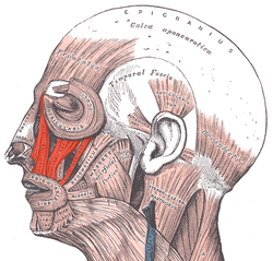
The levator labii superioris alaeque nasi muscle is, translated from Latin, the "lifter of both the upper lip and of the wing of the nose". The muscle is attached to the upper frontal process of the maxilla and inserts into the skin of the lateral part of the nostril and upper lip. At 44 characters, its name is longer than any other muscle's.

A snarl is a sound, often a growl or vicious utterance, often accompanied by a facial expression, where the upper lip is raised, and the nostrils widen, generally indicating hate, anger or pain. In addition to humans, other mammals including monkeys, rabbits and dogs snarl, often to warn others of their potential bite. In humans, snarling uses the levator labii superioris alaeque nasi muscle. The threatening vocalizations of snarling are often accompanied by or used synonymously with threatening facial expressions.

In the human skull, the zygomatic bone, also called cheekbone or malar bone, is a paired irregular bone, situated at the upper and lateral part of the face and forming part of the lateral wall and floor of the orbit, of the temporal fossa and the infratemporal fossa. It presents a malar and a temporal surface; four processes, and four borders.
In human anatomy, the orbicularis oris muscle is a complex of muscles in the lips that encircles the mouth. It is not a true sphincter, as was once thought, as it is actually composed of four independent quadrants that interlace and give only an appearance of circularity.

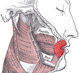
In anatomy, the masseter is one of the muscles of mastication. Found only in mammals, it is particularly powerful in herbivores to facilitate chewing of plant matter. The most obvious muscle of mastication is the masseter muscle, since it is the most superficial and one of the strongest.

The lips are a horizontal pair of soft appendages attached to the jaws and are the most visible part of the mouth of many animals, including humans. Vertebrate lips are soft, movable and serve to facilitate the ingestion of food and the articulation of sound and speech. Human lips are also a somatosensory organ, and can be an erogenous zone when used in kissing and other acts of intimacy.

The zygomaticus minor muscle is a muscle of facial expression. It originates from the zygomatic bone, lateral to the rest of the levator labii superioris muscle, and inserts into the outer part of the upper lip. It draws the upper lip backward, upward, and outward and is used in smiling. It is innervated by the facial nerve (VII).
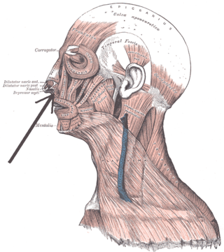
The depressor septi nasi muscle is a muscle of the face. It connects the incisive fossa of the maxilla and the orbicularis oris muscle to the nasal septum of the nose. It draws the ala of the nose downwards, reducing the size of the nostrils.

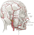
The facial artery is a branch of the external carotid artery that supplies structures of the superficial face.

The infraorbital artery is a small artery in the head that arises from the maxillary artery and passes through the inferior orbital fissure to enter the orbit, then passes forward along the floor of the orbit, finally exiting the orbit through the infraorbital foramen to reach the face.

The infraorbital nerve is a branch of the maxillary nerve. It arises in the pterygopalatine fossa. It passes through the inferior orbital fissure to enter the orbit. It travels through the orbit, then enters and traverses the infraorbital canal, exiting the canal at the infraorbital foramen to reach the face. It provides sensory innervation to the skin and mucous membranes around the middle of the face.

The angular artery is an artery of the face. It is the terminal part of the facial artery. It ascends to the medial angle of the eye's orbit. It is accompanied by the angular vein. It ends by anastomosing with the dorsal nasal branch of the ophthalmic artery. It supplies the lacrimal sac, the orbicularis oculi muscle, and the outer side of the nose.

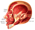
The facial muscles are a group of striated skeletal muscles supplied by the facial nerve that, among other things, control facial expression. These muscles are also called mimetic muscles. They are only found in mammals, although they derive from neural crest cells found in all vertebrates. They are the only muscles that attach to the dermis.

The human nose is the first organ of the respiratory system. It is also the principal organ in the olfactory system. The shape of the nose is determined by the nasal bones and the nasal cartilages, including the nasal septum, which separates the nostrils and divides the nasal cavity into two.

The infraorbital margin is the lower margin of the eye socket.

The following outline is provided as an overview of and topical guide to human anatomy:
Levator muscle can refer to:

The canine space, is a fascial space of the head and neck. It is a thin potential space on the face, and is paired on either side. It is located between the levator anguli oris muscle inferiorly and the levator labii superioris muscle superiorly. The term is derived from the fact that the space is in the region of the canine fossa, and that infections originating from the maxillary canine tooth may spread to involve the space. Infra-orbital is derived from infra- meaning below and orbit which refers to the eye socket.

Gummy smile, also known as excessive gingival display, is a smile that shows gum under the upper lip. It is a common clinical condition, which can be caused by an abnormal dental eruption, hyperfunction of the upper lip elevator muscle, excessive vertical growth of the maxilla bone, over-eruption of the maxillary anterior teeth, or a combination of the above described factors. Several treatment options have been proposed to enhance the smile display and to reduce the gingival exposure.