| Nasalis muscle | |
|---|---|
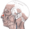
 The superior transverse part and inferior alar part of the nasalis muscle | |
| Details | |
| Origin | Maxilla |
| Insertion | Nasal bone |
| Artery | Superior labial artery |
| Nerve | Buccal branch of the facial nerve |
| Actions | Compresses bridge of nose, depresses tip of nose, elevates corners of nostrils |
| Identifiers | |
| Latin | musculus nasalis |
| TA98 | A04.1.03.009 |
| TA2 | 2062 |
| FMA | 46770 |
| Anatomical terms of muscle | |
The nasalis muscle is a sphincter-like muscle of the nose. It has a transverse part and an alar part. It compresses the nasal cartilages, and can "flare" the nostrils. It can be used to test the facial nerve (VII), which supplies it.