| Procerus muscle | |
|---|---|
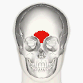
 Muscles of the head, face, and neck (Procerus visible at upper left, at top of nose) | |
| Details | |
| Origin | From fascia over the lower part of the nasal bone |
| Insertion | Into the skin of the lower part of the forehead between the eyebrows |
| Artery | Facial artery |
| Nerve | Temporal branch of the facial nerve |
| Actions | Draws down the medial angle of the eyebrow giving expressions of frowning |
| Identifiers | |
| Latin | musculus procerus, pyramidalis nasi, depressor glabellae |
| TA98 | A04.1.03.008 |
| TA2 | 2061 |
| FMA | 46769 |
| Anatomical terms of muscle | |
The procerus muscle (or pyramidalis nasi) is a small pyramidal muscle in the glabella. It is involved in facial expressions such as frowning and those associated with attentional control, and it indirectly helps shield the eyes from bright light. Because it contributes to wrinkle formation on the nasal bridge, it is often targeted in non-surgical facial rejuvenation treatments, such as botulinum toxin injections. [1] : 558 Procerus is Latin, meaning tall or extended.