Anterior auricular muscle

Last updated
Anterior auricular muscle
Sobo 1909 260 - Auricularis anterior.png
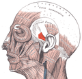
Face and neck muscles. Anterior auricular muscle shown in red.
Gray906 - Anterior auricular muscle.png
The muscles of the auricula. Anterior auricular is at right (indicated by the red arrow).
Details
Origin temporal fascia
Insertion major helix (ear)
Artery posterior auricular artery
Nerve temporal branch of facial nerve
Actions pulls ear forward
Identifiers
Latin musculus auricularis anterior
TA98 A04.1.03.020
TA2 2089
FMA 46856
Anatomical terms of muscle

The anterior auricular muscle, the smallest of the three auricular muscles, is thin and fan-shaped, and its fibers are pale and indistinct. It arises from the lateral edge of the epicranial aponeurosis, and its fibers converge to be inserted into a projection on the front of the helix.

Contents

Structure

The anterior auricular muscle arises from the lateral edge of the epicranial aponeurosis. [1] It inserts into a projection on the front of the helix. [1]

Nerve supply

The anterior auricular muscle is supplied is supplied by the temporal branch of the facial nerve (VII). [2] [3] It may also receive some small branches from the auriculotemporal nerve, a branch of the mandibular nerve, itself a branch of the trigeminal nerve (V). [4]

Relations

The anterior auricular muscle is the smallest of the three auricular muscles. [1]

The superficial temporal artery, a branch of the external carotid artery, travels underneath the anterior auricular muscle to supply the auricle of the outer ear. [1]

Function

The anterior auricular muscle draws the auricle of the outer ear upwards and forwards. [1] This is a very subtle movement in most people, although some people can wiggle their ears. [1]

See also

Additional images

Related Research Articles

<span class="mw-page-title-main">Glossopharyngeal nerve</span> Cranial nerve IX, for the tongue and pharynx

The glossopharyngeal nerve, also known as the ninth cranial nerve, cranial nerve IX, or simply CN IX, is a cranial nerve that exits the brainstem from the sides of the upper medulla, just anterior to the vagus nerve. Being a mixed nerve (sensorimotor), it carries afferent sensory and efferent motor information. The motor division of the glossopharyngeal nerve is derived from the basal plate of the embryonic medulla oblongata, whereas the sensory division originates from the cranial neural crest.

<span class="mw-page-title-main">Lesser occipital nerve</span> Nerve of the head and neck

The lesser occipital nerve is a cutaneous spinal nerve of the cervical plexus. It arises from second cervical (spinal) nerve (C2). It innervates the skin of the back of the upper neck and of the scalp posterior to the ear.

<span class="mw-page-title-main">Great auricular nerve</span> Cutaneous nerve of the head

The great auricular nerve is a cutaneous (sensory) nerve of the head. It originates from the second and third cervical (spinal) nerves (C2-C3) of the cervical plexus. It provides sensory innervation to the skin over the parotid gland and the mastoid process, parts of the outer ear, and to the parotid gland and its fascia.

<span class="mw-page-title-main">Cervical plexus</span> Network of nerves in the neck

The cervical plexus is a nerve plexus of the anterior rami of the first four cervical spinal nerves C1-C4. The cervical plexus provides motor innervation to some muscles of the neck, and the diaphragm; it provides sensory innervation to parts of the head, neck, and chest.

<span class="mw-page-title-main">Facial artery</span>

The facial artery is a branch of the external carotid artery that supplies structures of the superficial face.

<span class="mw-page-title-main">Foramen spinosum</span> Hole in the sphenoid bone of the skull

The foramen spinosum is a small open hole in the greater wing of the sphenoid bone that gives passage to the middle meningeal artery and vein, and the meningeal branch of the mandibular nerve.

<span class="mw-page-title-main">Occipitofrontalis muscle</span> Facial muscle helping to create facial expressions

The occipitofrontalis muscle is a muscle which covers parts of the skull. It consists of two parts or bellies: the occipital belly, near the occipital bone, and the frontal belly, near the frontal bone. It is supplied by the supraorbital artery, the supratrochlear artery, and the occipital artery. It is innervated by the facial nerve. In humans, the occipitofrontalis helps to create facial expressions.

<span class="mw-page-title-main">Foramen rotundum</span> Hole in the sphenoid bone of the skull

The foramen rotundum is a circular hole in the sphenoid bone of the skull. It connects the middle cranial fossa and the pterygopalatine fossa. It allows for the passage of the maxillary nerve (V2), a branch of the trigeminal nerve.

<span class="mw-page-title-main">Thyrohyoid membrane</span> Elastic membrane in the larynx

The thyrohyoid membrane is a broad, fibro-elastic sheet of the larynx. It connects the upper border of the thyroid cartilage to the hyoid bone.

<span class="mw-page-title-main">Supratrochlear nerve</span> Nerve of the forehead

The supratrochlear nerve is a branch of the frontal nerve, itself a branch of the ophthalmic nerve (CN V1) from the trigeminal nerve (CN V). It provides sensory innervation to the skin of the forehead and the upper eyelid.

<span class="mw-page-title-main">Posterior auricular artery</span> Small artery

The posterior auricular artery is a small artery that arises from the external carotid artery. It ascends along the side of the head. It supplies several muscles of the neck and several structures of the head.

<span class="mw-page-title-main">Retromandibular vein</span> Major face vein

The retromandibular vein is a major vein of the face. It is formed within the parotid gland by the confluence of the maxillary vein, and superficial temporal vein. It descends in the gland and splits into two branches upon emerging from the gland. Its anterior branch then joins the (anterior) facial vein forming the common facial vein, while its posterior branch joins the posterior auricular vein forming the external jugular vein.

<span class="mw-page-title-main">Mastoid part of the temporal bone</span> Back part of the sides of the skull base

The mastoid part of the temporal bone is the posterior (back) part of the temporal bone, one of the bones of the skull. Its rough surface gives attachment to various muscles and it has openings for blood vessels. From its borders, the mastoid part articulates with two other bones.

<span class="mw-page-title-main">Tympanic nerve</span>

The tympanic nerve is a branch of the glossopharyngeal nerve found near the ear. It gives sensation to the middle ear, the Eustachian tube, the parotid gland, and mastoid air cells. It gives parasympathetic to supply to the parotid gland via the otic ganglion and the auriculotemporal nerve.

<span class="mw-page-title-main">Posterior ethmoidal nerve</span> Nerve of the orbit around the eye

The posterior ethmoidal nerve is a nerve of the head. It is a branch of the nasociliary nerve (itself a branch of the ophthalmic nerve (CN V1)). It provides sensory innervation to the sphenoid sinus and ethmoid sinus, and part of the dura mater in the anterior cranial fossa.

<span class="mw-page-title-main">Medial pterygoid nerve</span> Nerve of the head

The medial pterygoid nerve (nerve to medial pterygoid, or internal pterygoid nerve) is a nerve of the head. It is a branch of the mandibular nerve (CN V3). It supplies the medial pterygoid muscle, the tensor veli palatini muscle, and the tensor tympani muscle.

<span class="mw-page-title-main">Posterior auricular nerve</span> Nerve of the head

The posterior auricular nerve is a nerve of the head. It is a branch of the facial nerve. It communicates with branches from the vagus nerve, the great auricular nerve, and the lesser occipital nerve. Its auricular branch supplies the posterior auricular muscle, the intrinsic muscles of the auricle, and gives sensation to the auricle. Its occipital branch supplies the occipitalis muscle.

<span class="mw-page-title-main">Zygomatic branches of the facial nerve</span> Nerves of the face

The zygomatic branches of the facial nerve (malar branches) are nerves of the face. They run across the zygomatic bone to the lateral angle of the orbit. Here, they supply the orbicularis oculi muscle, and join with filaments from the lacrimal nerve and the zygomaticofacial branch of the maxillary nerve (CN V2).

<span class="mw-page-title-main">Posterior auricular muscle</span> Muscle that pulls the ear upward and backward

The posterior auricular muscle is a muscle behind the auricle of the outer ear. It arises from the mastoid part of the temporal bone, and inserts into the lower part of the cranial surface of the auricle of the outer ear. It draws the auricle backwards, usually a very slight effect.

<span class="mw-page-title-main">Superior auricular muscle</span> Muscle that draws the auricle of the outer ear upwards

The superior auricular muscle is a muscle above the auricle of the outer ear. It originates from the epicranial aponeurosis, and inserts into the upper part of the medial surface of the auricle. It draws the auricle upwards.

References

PD-icon.svgThis article incorporates text in the public domain from page 1035 of the 20th edition of Gray's Anatomy (1918)

  1. 1 2 3 4 5 6 Niamtu, Joseph (2018). "8 - Cosmetic Otoplasty and Related Ear Surgery". Cosmetic Facial Surgery (2nd ed.). Edinburgh: Elsevier. pp. 473–532. doi:10.1016/B978-0-323-39393-5.00008-X. ISBN   978-0-323-39402-4. OCLC   976037123.
  2. Barral, Jean Pierre; Croibier, Alain (2009). "19 - Facial nerve". Manual therapy for the cranial nerves. Edinburgh: Churchill Livingstone / Elsevier. pp. 153–166. doi:10.1016/B978-0-7020-3100-7.50022-7. ISBN   978-0-7020-3736-8. OCLC   460904284.
  3. Rea, Paul (2016). "2 - Head". Essential clinically applied anatomy of the peripheral nervous system in the head and neck. Amsterdam: Academic Press, Elsevier Science. pp. 21–130. doi:10.1016/B978-0-12-803633-4.00002-8. ISBN   0-12-803664-8. OCLC   939866688.
  4. Barral, Jean-Pierre; Croibier, Alain (2009). "25 - Ear". Manual Therapy for the Cranial Nerves. Edinburgh: Churchill Livingstone / Elsevier. pp. 227–238. doi:10.1016/B978-0-7020-3100-7.50028-8. ISBN   978-0-7020-3736-8. OCLC   460904284.