Epicranial aponeurosis

Last updated
Epicranial aponeurosis
1106 Side Views of the Muscles of Facial Expressions numbered.jpg
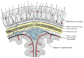
Muscles of the head, face, and neck. (Epicranial aponeurosis visible at top labeled 1.)
Details
System skeletal
Identifiers
Latin galea aponeurotica,
aponeurosis epicranialis,
aponeurosis epicrania
TA98 A04.1.03.007
TA2 2059
FMA 46768
Anatomical terminology

The epicranial aponeurosis (aponeurosis epicranialis, galea aponeurotica) is an aponeurosis (a tough layer of dense fibrous tissue). It covers the upper part of the skull in humans and many other animals.

Contents

Structure

In humans, the epicranial aponeurosis originates from the external occipital protuberance and highest nuchal lines of the occipital bone. [1] It merges with the occipitofrontalis muscle. In front, it forms a short and narrow prolongation between its union with the frontalis muscle (the frontal part of the occipitofrontalis muscle).

On either side, the epicranial aponeurosis attaches to the anterior auricular muscles and the superior auricular muscles. Here it is less aponeurotic, and is continued over the temporal fascia to the zygomatic arch as a layer of laminated areolar tissue.

It is closely connected to the integument by the firm, dense, fibro-fatty layer which forms the superficial fascia of the scalp. It is attached to the pericranium by loose cellular tissue, which allows the aponeurosis, carrying with it the integument, to move through a considerable distance.

Clinical significance

Subgaleal haemorrhage is defined as bleeding between the epicranial aponeurosis and the skull. [2] Conservative management is usually appropriate for these, as there is little risk of further damage to surrounding structures. [2]

History

The epicranial aponeurosis is also known as the aponeurosis epicranialis (from Latin),[ citation needed ] and the galea aponeurotica. [2]

Additional images

See also

Related Research Articles

<span class="mw-page-title-main">Lesser occipital nerve</span> Nerve of the head and neck

The lesser occipital nerve is a cutaneous spinal nerve of the cervical plexus. It arises from second cervical (spinal) nerve (C2). It innervates the skin of the back of the upper neck and of the scalp posterior to the ear.

<span class="mw-page-title-main">Parietal bone</span> Bone in the human skull which, when joined together, forms the sides and roof of the cranium

The parietal bones are two bones in the skull which, when joined at a fibrous joint, form the sides and roof of the cranium. In humans, each bone is roughly quadrilateral in form, and has two surfaces, four borders, and four angles. It is named from the Latin paries (-ietis), wall.

<span class="mw-page-title-main">Scalp</span> Anatomical area bordered by the face at the front, and by the neck at the sides and back

The scalp is the anatomical area bordered by the face at the front, and by the neck at the sides and back.

<span class="mw-page-title-main">Aponeurosis</span> Tissue which connects muscles to other organs

An aponeurosis is a flattened tendon by which muscle attaches to bone or fascia. Aponeuroses exhibit an ordered arrangement of collagen fibres, thus attaining high tensile strength in a particular direction while being vulnerable to tensional or shear forces in other directions. They have a shiny, whitish-silvery color, are histologically similar to tendons, and are very sparingly supplied with blood vessels and nerves. When dissected, aponeuroses are papery and peel off by sections. The primary regions with thick aponeuroses are in the ventral abdominal region, the dorsal lumbar region, the ventriculus in birds, and the palmar (palms) and plantar (soles) regions.

<span class="mw-page-title-main">Platysma muscle</span> Human neck muscle

The platysma muscle is a superficial muscle of the human neck that overlaps the sternocleidomastoid. It covers the anterior surface of the neck superficially. When it contracts, it produces a slight wrinkling of the neck, and a "bowstring" effect on either side of the neck.

<span class="mw-page-title-main">Frontalis muscle</span>

The frontalis muscle is a muscle which covers parts of the forehead of the skull. Some sources consider the frontalis muscle to be a distinct muscle. However, Terminologia Anatomica currently classifies it as part of the occipitofrontalis muscle along with the occipitalis muscle.

<span class="mw-page-title-main">Occipitalis muscle</span>

The occipitalis muscle is a muscle which covers parts of the skull. Some sources consider the occipital muscle to be a distinct muscle. However, Terminologia Anatomica currently classifies it as part of the occipitofrontalis muscle along with the frontalis muscle.

The epicranium is the medical term for the collection of structures covering the cranium. It consists of the muscles, aponeurosis, and skin.

<span class="mw-page-title-main">Superficial temporal artery</span> Major artery of the head

In human anatomy, the superficial temporal artery is a major artery of the head. It arises from the external carotid artery when it splits into the superficial temporal artery and maxillary artery.

<span class="mw-page-title-main">Occipitofrontalis muscle</span> Facial muscle helping to create facial expressions

The occipitofrontalis muscle is a muscle which covers parts of the skull. It consists of two parts or bellies: the occipital belly, near the occipital bone, and the frontal belly, near the frontal bone. It is supplied by the supraorbital artery, the supratrochlear artery, and the occipital artery. It is innervated by the facial nerve. In humans, the occipitofrontalis helps to create facial expressions.

<span class="mw-page-title-main">Occipital artery</span>

The occipital artery is a branch of the external carotid artery that provides arterial supply to the back of the scalp, sternocleidomastoid muscles, and deep muscles of the back and neck.

<span class="mw-page-title-main">Nuchal lines</span> Part of the skulls occipital bone

The nuchal lines are four curved lines on the external surface of the occipital bone:

<span class="mw-page-title-main">Occipital vein</span> Vein of the scalp

The occipital vein is a vein of the scalp. It originates from a plexus around the external occipital protuberance and superior nuchal line to the back part of the vertex of the skull. It usually drains into the internal jugular vein, but may also drain into the posterior auricular vein. It drains part of the scalp.

<span class="mw-page-title-main">Transversalis fascia</span> Aponeurosis between the transverse abdominal muscle and the extraperitoneal fat

The transversalis fascia is the fascial lining of the anterolateral abdominal wall situated between the inner surface of the transverse abdominal muscle, and the preperitoneal fascia. It is directly continuous with the iliac fascia, the internal spermatic fascia, and pelvic fascia.

<span class="mw-page-title-main">Posterior auricular nerve</span> Nerve of the head

The posterior auricular nerve is a nerve of the head. It is a branch of the facial nerve. It communicates with branches from the vagus nerve, the great auricular nerve, and the lesser occipital nerve. Its auricular branch supplies the posterior auricular muscle, the intrinsic muscles of the auricle, and gives sensation to the auricle. Its occipital branch supplies the occipitalis muscle.

The cremasteric fascia is a fascia in the scrotum. As the cremaster descends, it forms a series of loops which differ in thickness and length in different subjects. At the upper part of the cord the loops are short, but they become in succession longer and longer, the longest reaching down as low as the testis, where a few are inserted into the tunica vaginalis. These loops are united together by areolar tissue, and form a thin covering over the cord and testis, the cremasteric fascia.

<span class="mw-page-title-main">External spermatic fascia</span>

The external spermatic fascia is a thin membrane, prolonged downward around the surface of the spermatic cord and testis. It is separated from the dartos tunic by loose areolar tissue. It is occasionally referred to as 'Le Fascia de Webster' after an anatomist who once described it.

<span class="mw-page-title-main">Superior auricular muscle</span> Muscle that draws the auricle of the outer ear upwards

The superior auricular muscle is a muscle above the auricle of the outer ear. It originates from the epicranial aponeurosis, and inserts into the upper part of the medial surface of the auricle. It draws the auricle upwards.

<span class="mw-page-title-main">Anterior auricular muscle</span> Muscle connectingthe epicranial aponeurosis to the helix of the ear

The anterior auricular muscle, the smallest of the three auricular muscles, is thin and fan-shaped, and its fibers are pale and indistinct. It arises from the lateral edge of the epicranial aponeurosis, and its fibers converge to be inserted into a projection on the front of the helix.

Scalp reconstruction is a surgical procedure for people with scalp defects. Scalp defects may be partial or full thickness and can be congenital or acquired. Because not all layers of the scalp are elastic and the scalp has a convex shape, the use of primary closure is limited. Sometimes the easiest way of closing the wound may not be the ideal or best way. The choice for a reconstruction depends on multiple factors, such as the defect itself, the patient characteristics and surgeon preference.

References

PD-icon.svgThis article incorporates text in the public domain from page 380 of the 20th edition of Gray's Anatomy (1918)

  1. Cunningham, Craig; Scheuer, Louise; Black, Sue (2016). "5 - The Skull". Developmental juvenile osteology (2nd ed.). Amsterdam: Academic Press. pp. 43–148. doi:10.1016/B978-0-12-382106-5.00005-0. ISBN   978-0-12-382107-2. OCLC   956277358.
  2. 1 2 3 Stewart, Christopher C. (2007). "143 - Neurosurgical Issues". Comprehensive Pediatric Hospital Medicine. Philadelphia: Mosby. pp. 908–914. doi:10.1016/B978-032303004-5.50147-2. ISBN   978-0-323-07040-9. OCLC   324998103.