Superior auricular muscle | |
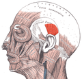
---|---|
Details | |
Origin | temporal fascia |
Insertion | above the auricle of the outer ear |
Artery | posterior auricular artery |
Nerve | branches to auricular muscle from posterior auricular nerve of facial nerve (cranial nerve VII) |
Actions | pulls ear upward |
Identifiers | |
Latin | musculus auricularis superior |
TA98 | A04.1.03.021 |
TA2 | 2090 |
FMA | 46855 |
Anatomical terms of muscle |
The superior auricular muscle is a muscle above the auricle of the outer ear. It originates from the epicranial aponeurosis, and inserts into the upper part of the medial surface of the auricle. It draws the auricle upwards.
The superior auricular muscle originates from the epicranial aponeurosis. [1] Its fibres converge to be inserted by a thin, flattened tendon into the upper part of the medial surface of the auricle of the outer ear. [1]
It is the largest of the three auriculares muscles. [1] It is thin and fan-shaped.
The superior auricular muscle is supplied by the temporal branch of the facial nerve (VII). [1] [2]
Wikimedia Commons has media related to Auricularis superior muscles . |
The outer ear, external ear, or auris externa is the external part of the ear, which consists of the auricle and the ear canal . It gathers sound energy and focuses it on the eardrum.
The trapezius is a large paired trapezoid-shaped surface muscle that extends longitudinally from the occipital bone to the lower thoracic vertebrae of the spine and laterally to the spine of the scapula. It moves the scapula and supports the arm.
The sartorius muscle is the longest muscle in the human body. It is a long, thin, superficial muscle that runs down the length of the thigh in the anterior compartment.
The sternocleidomastoid muscle is one of the largest and most superficial cervical muscles. The primary actions of the muscle are rotation of the head to the opposite side and flexion of the neck. The sternocleidomastoid is innervated by the accessory nerve.
Pectoralis minor muscle is a thin, triangular muscle, situated at the upper part of the chest, beneath the pectoralis major in the human body.
The cervical plexus is a plexus of the anterior rami of the first four cervical spinal nerves which arise from C1 to C4 cervical segment in the neck. They are located laterally to the transverse processes between prevertebral muscles from the medial side and vertebral from lateral side. There is anastomosis with accessory nerve, hypoglossal nerve and sympathetic trunk.
The quadriceps femoris muscle is a large muscle group that includes the four prevailing muscles on the front of the thigh. It is the great extensor muscle of the knee, forming a large fleshy mass which covers the front and sides of the femur. The name derives from Latin four-headed muscle of the femur.
The levator palpebrae superioris is the muscle in the orbit that elevates the upper eyelid.
The internal obturator muscle or obturator internus muscle originates on the medial surface of the obturator membrane, the ischium near the membrane, and the rim of the pubis.
The stylohyoid muscle is a slender muscle, lying anterior and superior of the posterior belly of the digastric muscle. It is one of the suprahyoid muscles. It shares this muscle's innervation by the facial nerve, and functions to draw the hyoid bone backwards and elevate the tongue. Its origin is the styloid process of the temporal bone. It inserts on the body of the hyoid.
The epicranial aponeurosis is an aponeurosis. It covers the upper part of the skull in humans and many other animals.
The abdominal external oblique muscle is the largest and outermost of the three flat abdominal muscles of the lateral anterior abdomen.
The semimembranosus muscle is the most medial of the three hamstring muscles in the thigh. It is so named because it has a flat tendon of origin. It lies posteromedially in the thigh, deep to the semitendinosus muscle. It extends the hip joint and flexes the knee joint.
The occipitofrontalis muscle is a muscle which covers parts of the skull. It consists of two parts or bellies: the occipital belly, near the occipital bone, and the frontal belly, near the frontal bone. It is supplied by the supraorbital artery, the supratrochlear artery, and the occipital artery. It is innervated by the facial nerve. In humans, the occipitofrontalis helps to create facial expressions.
The nuchal lines are four curved lines on the external surface of the occipital bone:
The adductor canal, also known as the subsartorial canal or Hunter’s canal, is an aponeurotic tunnel in the middle third of the thigh. It extends from the apex of the femoral triangle to the adductor hiatus.
The retromandibular vein is a major vein of the face.
The mastoid part of the temporal bone is the posterior (back) part of the temporal bone, one of the bones of the skull. Its rough surface gives attachment to various muscles and it has openings for blood vessels. From its borders, the mastoid part articulates with two other bones.
The posterior auricular muscle is a muscle behind the auricle of the outer ear. It arises from the mastoid part of the temporal bone, and inserts into the lower part of the cranial surface of the auricle of the outer ear. It draws the auricle backwards, usually a very slight effect.
The anterior auricular muscle, the smallest of the three auricular muscles, is thin and fan-shaped, and its fibers are pale and indistinct. It arises from the lateral edge of the epicranial aponeurosis, and its fibers converge to be inserted into a projection on the front of the helix.
This article incorporates text in the public domain from page 1035 of the 20th edition of Gray's Anatomy (1918)