Levator palpebrae superioris muscle

Last updated
Levator palpebrae superioris
Eyemuscles.png
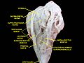
Rectus muscles:
2 = superior, 3 = inferior, 4 = medial, 5 = lateral
Oblique muscles: 6 = superior, 8 = inferior
Other muscle: 9 = levator palpebrae superioris
Other structures: 1 = Annulus of Zinn, 7 = Trochlea, 10 = Superior tarsus, 11 = Sclera, 12 = Optic nerve
Gray891.png
The levator palebrae superioris can be seen here, travelling above the superior rectus muscle, and ending at the upper eyelid.
Details
Origin Inferior surface of lesser wing of sphenoid
Insertion Superior tarsal plate and skin of upper eyelid
Artery Muscular branches of ophthalmic artery and supraorbital artery
Nerve Superior division of oculomotor nerve
Actions Elevation of upper eyelid
Antagonist Palpebral part of orbicularis oculi muscle
Identifiers
Latin musculus levator palpebrae superioris
TA98 A15.2.07.020
TA2 2052
FMA 49041
Anatomical terms of muscle

The levator palpebrae superioris (Latin : elevating muscle of upper eyelid) is the muscle in the orbit that elevates the upper eyelid. [1] [2]

Contents

Structure

The levator palpebrae superioris originates from inferior surface of the lesser wing of the sphenoid bone, just above the optic foramen. It broadens and decreases in thickness (becomes thinner) and becomes the levator aponeurosis. This portion inserts on the skin of the upper eyelid, as well as the superior tarsal plate. It is a skeletal muscle. The superior tarsal muscle, a smooth muscle, is attached to the levator palpebrae superioris, and inserts on the superior tarsal plate as well.

Blood supply

The levator palebrae superioris receives its blood supply from branches of the ophthalmic artery, specifically, muscular branches and the supraorbital artery. Blood is drained into the superior ophthalmic vein.

Nerve supply

The levator palpebrae superioris receives motor innervation from the superior division of the oculomotor nerve. [1] [2] [3] The smooth muscle that originates from its undersurface, called the superior tarsal muscle is innervated by postganglionic sympathetic axons from the superior cervical ganglion. [2]

Function

The levator palpebrae superioris elevates the upper eyelid. [1] [2]

Clinical significance

Damage to this muscle or its innervation can cause ptosis, which is drooping of the eyelid. [4] [5] Lesions in CN III can cause ptosis, [5] because without stimulation from the oculomotor nerve the levator palpebrae cannot oppose the force of gravity, and the eyelid droops.

Ptosis can also result from damage to the adjoining superior tarsal muscle or its sympathetic innervation. Such damage to the sympathetic supply occurs in Horner's syndrome and presents as a partial ptosis. It is important to distinguish between these two very different causes of ptosis. This can usually be done clinically without issue, as each type of ptosis is accompanied by other distinct clinical findings.

The ptosis seen in paralysis of the levator palpebrae superioris is usually more pronounced than that seen due to paralysis of the superior tarsal muscle.

Additional images

See also

Related Research Articles

<span class="mw-page-title-main">Danger triangle of the face</span> Area of the face with blood supply that connects indirectly to the brain

The danger triangle of the face consists of the area from the corners of the mouth to the bridge of the nose, including the nose and maxilla. Due to the special nature of the blood supply to the human nose and surrounding area, it is possible, albeit extremely unlikely, for retrograde infection from the nasal area to spread to the brain, causing cavernous sinus thrombosis, meningitis, or brain abscess.

<span class="mw-page-title-main">Oculomotor nerve</span> Cranial nerve III, for eye movements

The oculomotor nerve, also known as the third cranial nerve, cranial nerve III, or simply CN III, is a cranial nerve that enters the orbit through the superior orbital fissure and innervates extraocular muscles that enable most movements of the eye and that raise the eyelid. The nerve also contains fibers that innervate the intrinsic eye muscles that enable pupillary constriction and accommodation. The oculomotor nerve is derived from the basal plate of the embryonic midbrain. Cranial nerves IV and VI also participate in control of eye movement.

<span class="mw-page-title-main">Supraorbital nerve</span> Branch of the frontal nerve

The supraorbital nerve is one of two branches of the frontal nerve, itself a branch of the ophthalmic nerve. The other branch of the frontal nerve is the supratrochlear nerve.

<span class="mw-page-title-main">Superior rectus muscle</span> Extraocular muscle that elevates the eye

The superior rectus muscle is a muscle in the orbit. It is one of the extraocular muscles. It is innervated by the superior division of the oculomotor nerve (III). In the primary position, its primary function is elevation, although it also contributes to intorsion and adduction. It is associated with a number of medical conditions, and may be weak, paralysed, overreactive, or even congenitally absent in some people.

<span class="mw-page-title-main">Extraocular muscles</span> Seven extrinsic muscles of the human eye

The extraocular muscles, or extrinsic ocular muscles, are the seven extrinsic muscles of the human eye. Six of the extraocular muscles, the four recti muscles, and the superior and inferior oblique muscles, control movement of the eye and the other muscle, the levator palpebrae superioris, controls eyelid elevation. The actions of the six muscles responsible for eye movement depend on the position of the eye at the time of muscle contraction.

<span class="mw-page-title-main">Superior orbital fissure</span> Foramen in the skull allowing for passage of cranial nerves

The superior orbital fissure is a foramen or cleft of the skull between the lesser and greater wings of the sphenoid bone. It gives passage to multiple structures, including the oculomotor nerve, trochlear nerve, ophthalmic nerve, abducens nerve, ophthalmic veins, and sympathetic fibres from the cavernous plexus.

<span class="mw-page-title-main">Ophthalmic artery</span> Artery of the head

The ophthalmic artery (OA) is an artery of the head. It is the first branch of the internal carotid artery distal to the cavernous sinus. Branches of the ophthalmic artery supply all the structures in the orbit around the eye, as well as some structures in the nose, face, and meninges. Occlusion of the ophthalmic artery or its branches can produce sight-threatening conditions.

<span class="mw-page-title-main">Ophthalmic nerve</span> Sensory nerve of the face

The ophthalmic nerve (CN V1) is a sensory nerve of the head. It is one of three divisions of the trigeminal nerve (CN V), a cranial nerve. It has three major branches which provide sensory innervation to the eye, and the skin of the upper face and anterior scalp, as well as other structures of the head.

<span class="mw-page-title-main">Nasociliary nerve</span> Branch of the ophthalmic nerve

The nasociliary nerve is a branch of the ophthalmic nerve (CN V1) (which is in turn a branch of the trigeminal nerve (CN V)). It is intermediate in size between the other two branches of the ophthalmic nerve, the frontal nerve and lacrimal nerve.

<span class="mw-page-title-main">Tarsus (eyelids)</span>

The tarsi or tarsal plates are two comparatively thick, elongated plates of dense connective tissue, about 10 mm (0.39 in) in length for the upper eyelid and 5 mm for the lower eyelid; one is found in each eyelid, and contributes to its form and support. They are located directly above the lid margins. The tarsus has a lower and upper part making up the palpebrae.

<span class="mw-page-title-main">Frontal nerve</span> Nerve supplying the forehead, frontal sinus, and upper eyelid

The frontal nerve is the largest branch of the ophthalmic nerve (V1), itself a branch of the trigeminal nerve (CN V). It supplies sensation to the skin of the forehead, the mucosa of the frontal sinus, and the skin of the upper eyelid. It may be affected by schwannoma.

<span class="mw-page-title-main">Supratrochlear nerve</span> Nerve of the forehead

The supratrochlear nerve is a branch of the frontal nerve, itself a branch of the ophthalmic nerve (CN V1) from the trigeminal nerve (CN V). It provides sensory innervation to the skin of the forehead and the upper eyelid.

The superior transverse ligament of the eye is a transverse ligament surrounding the levator palpebrae superioris muscle close to its partial implantation into the skin of the upper eyelid. The muscle also attaches to the superior tarsal plate and into orbital bone.

<span class="mw-page-title-main">Superior tarsal muscle</span>

The superior tarsal muscle is a smooth muscle adjoining the levator palpebrae superioris muscle that helps to raise the upper eyelid.

<span class="mw-page-title-main">Supraorbital artery</span>

The supraorbital artery is a branch of the ophthalmic artery. It passes anteriorly within the orbit to exit the orbit through the supraorbital foramen or notch alongside the supraorbital nerve, splitting into two terminal branches which go on to form anastomoses with arteries of the head.

<span class="mw-page-title-main">Orbital septum</span>

In anatomy, the orbital septum is a membranous sheet that acts as the anterior (frontal) boundary of the orbit. It extends from the orbital rims to the eyelids. It forms the fibrous portion of the eyelids.

<span class="mw-page-title-main">Ptosis (eyelid)</span> Drooping of the upper eyelid over the eye

Ptosis, also known as blepharoptosis, is a drooping or falling of the upper eyelid. This condition is sometimes called "lazy eye," but that term normally refers to the condition amblyopia. If severe enough and left untreated, the drooping eyelid can cause other conditions, such as amblyopia or astigmatism, so it is especially important to treat the disorder in children before it can interfere with vision development.

<span class="mw-page-title-main">Marcus Gunn phenomenon</span> Medical condition

Marcus Gunn phenomenon is an autosomal dominant condition with incomplete penetrance, in which nursing infants will have rhythmic upward jerking of their upper eyelid. This condition is characterized as a synkinesis: when two or more muscles that are independently innervated have either simultaneous or coordinated movements.

<span class="mw-page-title-main">Oculomotor nerve palsy</span> Medical condition

Oculomotor nerve palsy or oculomotor neuropathy is an eye condition resulting from damage to the third cranial nerve or a branch thereof. As the name suggests, the oculomotor nerve supplies the majority of the muscles controlling eye movements. Damage to this nerve will result in an inability to move the eye normally. The nerve also supplies the upper eyelid muscle and is accompanied by parasympathetic fibers innervating the muscles responsible for pupil constriction. The limitations of eye movement resulting from the condition are generally so severe that patients are often unable to maintain normal eye alignment when gazing straight ahead, leading to strabismus and, as a consequence, double vision (diplopia).

<span class="mw-page-title-main">Roots of the ciliary ganglion</span>

The ciliary ganglion is a parasympathetic ganglion located just behind the eye in the posterior orbit. Three types of axons enter the ciliary ganglion but only the preganglionic parasympathetic axons synapse there. The entering axons are arranged into three roots of the ciliary ganglion, which join enter the posterior surface of the ganglion.

References

  1. 1 2 3 Liu, Grant T. (2003-01-01), Samuels, Martin A.; Feske, Steven K. (eds.), "Chapter 6 - Disorders of the Eyes and Eyelids", Office Practice of Neurology (Second Edition), Philadelphia: Churchill Livingstone, pp. 35–69, doi:10.1016/b0-44-306557-8/50008-3, ISBN   978-0-443-06557-6 , retrieved 2020-11-11
  2. 1 2 3 4 Standring, Susan, ed. (2016). ""Extraocular muscles: levator palpebrae superioris"". Gray's anatomy: the anatomical basis of clinical practice (41st ed.). Philadelphia. p. 670. ISBN   9780702052309. OCLC   920806541.{{cite book}}: CS1 maint: location missing publisher (link)
  3. Jackson, Timothy L., ed. (2008-01-01), "Chapter 1 - OCULOPLASTICS", Moorfields Manual of Ophthalmology, Edinburgh: Mosby, pp. 1–54, doi:10.1016/b978-1-4160-2572-6.50006-x, ISBN   978-1-4160-2572-6, S2CID   241607885 , retrieved 2020-11-11
  4. Trobe, Jonathan D. (2008-01-01), Trobe, Jonathan D. (ed.), "Section 13 - Eyelid Disorders", Neuro-ophthalmology, Edinburgh: Mosby, pp. 229–239, doi: 10.1016/b978-0-323-04456-1.50016-9 , ISBN   978-0-323-04456-1
  5. 1 2 Hejtmancik, J. F.; Cabrera, P.; Chen, Y.; M’Hamdi, O.; Nickerson, J. M. (2017-01-01), Conn, P. Michael (ed.), "Chapter 19 - Vision", Conn's Translational Neuroscience, San Diego: Academic Press, pp. 399–438, doi:10.1016/b978-0-12-802381-5.00031-2, ISBN   978-0-12-802381-5 , retrieved 2020-11-11