In human anatomy, a hamstring is any one of the three posterior thigh muscles in between the hip and the knee. The hamstrings are susceptible to injury. In quadrupeds, the hamstring is the single large tendon found behind the knee or comparable area.

In anatomy, the orbit is the cavity or socket of the skull in which the eye and its appendages are situated. "Orbit" can refer to the bony socket, or it can also be used to imply the contents. In the adult human, the volume of the orbit is 30 millilitres, of which the eye occupies 6.5 ml. The orbital contents comprise the eye, the orbital and retrobulbar fascia, extraocular muscles, cranial nerves II, III, IV, V, and VI, blood vessels, fat, the lacrimal gland with its sac and duct, the eyelids, medial and lateral palpebral ligaments, check ligaments, the suspensory ligament, septum, ciliary ganglion and short ciliary nerves.
An eyelid is a thin fold of skin that covers and protects an eye. The levator palpebrae superioris muscle retracts the eyelid, exposing the cornea to the outside, giving vision. This can be either voluntarily or involuntarily. The human eyelid features a row of eyelashes along the eyelid margin, which serve to heighten the protection of the eye from dust and foreign debris, as well as from perspiration. "Palpebral" means relating to the eyelids. Its key function is to regularly spread the tears and other secretions on the eye surface to keep it moist, since the cornea must be continuously moist. They keep the eyes from drying out when asleep. Moreover, the blink reflex protects the eye from foreign bodies.

Canthus is either corner of the eye where the upper and lower eyelids meet. More specifically, the inner and outer canthi are, respectively, the medial and lateral ends/angles of the palpebral fissure.

The extraocular muscles, are the seven extrinsic muscles of the human eye. Six of the extraocular muscles control movement of the eye and the other muscle the levator palpebrae controls eyelid elevation. The actions of the six muscles responsible for eye movement depend on the position of the eye at the time of muscle contraction.

Blepharoplasty is the plastic surgery operation for correcting defects, deformities, and disfigurations of the eyelids; and for aesthetically modifying the eye region of the face. With the excision and the removal, or the repositioning of excess tissues, such as skin and adipocyte fat, and the reinforcement of the corresponding muscle and tendon tissues, the blepharoplasty procedure resolves functional and cosmetic problems of the periorbita, which is the area from the eyebrow to the upper portion of the cheek. The procedure is more common among women, who accounted for approximately 85% of blepharoplasty procedures in 2014 in the US and 88% of such procedures in the UK.

The orbicularis oculi is a muscle in the face that closes the eyelids. It arises from the nasal part of the frontal bone, from the frontal process of the maxilla in front of the lacrimal groove, and from the anterior surface and borders of a short fibrous band, the medial palpebral ligament.

The tarsi are two comparatively thick, elongated plates of dense connective tissue, about 10 mm (0.39 in) in length for the upper eyelid and 5 mm for the lower eyelid; one is found in each eyelid, and contributes to its form and support. They are located directly above the lid margins. The tarsus has a lower and upper part making up the palpebrae.
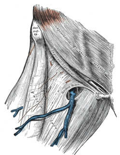
The fascia lata is the deep fascia of the thigh. It encloses the thigh muscles and forms the outer limit of the fascial compartments of thigh, which are internally separated by intermuscular septa. The fascia lata is thickened at its lateral side where it forms the iliotibial tract, a structure that runs to the tibia and serves as a site of muscle attachment.

The lacrimal sac or lachrymal sac is the upper dilated end of the nasolacrimal duct, and is lodged in a deep groove formed by the lacrimal bone and frontal process of the maxilla. It connects the lacrimal canaliculi, which drain tears from the eye's surface, and the nasolacrimal duct, which conveys this fluid into the nasal cavity. Lacrimal sac occlusion leads to dacryocystitis.
The anterior lacrimal crest is a bony projection on the frontal process of the maxilla. It creates the lateral margin of the lacrimal sac fossa and is continuous with the orbital margin. The medial palpebral ligament is attached to anterior lacrimal crest. It is an important structure to avoid damaging during rhinoplasty.
The medial palpebral ligament is a ligament of the face. It attaches to the frontal process of the maxilla, the lacrimal groove, and the tarsus of each eyelid. It has a superficial (anterior) and a deep (posterior) layer, with many surrounding attachments. It connects the medial canthus of each eyelid to the medial part of the orbit. It is a useful point of fixation during eyelid reconstructive surgery.
The superior tarsal muscle is a smooth muscle adjoining the levator palpebrae superioris muscle that helps to raise the upper eyelid.

Graves’ ophthalmopathy, also known as thyroid eye disease (TED), is an autoimmune inflammatory disorder of the orbit and periorbital tissues, characterized by upper eyelid retraction, lid lag, swelling, redness (erythema), conjunctivitis, and bulging eyes (exophthalmos). It occurs most commonly in individuals with Graves' disease, and less commonly in individuals with Hashimoto's thyroiditis, or in those who are euthyroid.

The Tenon capsule, also known as the fascial sheath of the eyeball or the fascia bulbi, is a thin membrane which envelops the eyeball from the optic nerve to the corneal limbus, separating it from the orbital fat and forming a socket in which it moves.

Orbital cellulitis is inflammation of eye tissues behind the orbital septum. It is most commonly caused by an acute spread of infection into the eye socket from either the adjacent sinuses or through the blood. It may also occur after trauma. When it affects the rear of the eye, it is known as retro-orbital cellulitis.

Periorbital cellulitis or preseptal cellulitis, is an inflammation and infection of the eyelid and portions of skin around the eye anterior to the orbital septum. It may be caused by breaks in the skin around the eye, and subsequent spread to the eyelid; infection of the sinuses around the nose (sinusitis); or from spread of an infection elsewhere through the blood.
Oculoplastics, or oculoplastic surgery, includes a wide variety of surgical procedures that deal with the orbit, eyelids, tear ducts, and the face. It also deals with the reconstruction of the eye and associated structures.
The lateral palpebral raphe is a ligamentous band near the eye. Its existence is contentious, and many sources describe it as the continuation of nearby muscles. It is formed from the lateral ends of the orbicularis oculi muscle. It connects the orbicularis orbis muscle, the frontosphenoidal process of the zygomatic bone, and the tarsi of the eyelids.

Medial knee injuries are the most common type of knee injury. The medial ligament complex of the knee is composed of the superficial medial collateral ligament (sMCL), deep medial collateral ligament (dMCL), and the posterior oblique ligament (POL). These ligaments have also been called the medial collateral ligament (MCL), tibial collateral ligament, mid-third capsular ligament, and oblique fibers of the sMCL, respectively. This complex is the major stabilizer of the medial knee. Injuries to the medial side of the knee are most commonly isolated to these ligaments. A thorough understanding of the anatomy and function of the medial knee structures, along with a detailed history and physical exam, are imperative to diagnosing and treating these injuries.
