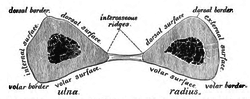
The ulna is a long bone found in the forearm that stretches from the elbow to the smallest finger, and when in anatomical position, is found on the medial side of the forearm. It runs parallel to the radius, the other long bone in the forearm, and is the larger and longer of the two.

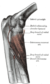
The radial nerve is a nerve in the human body that supplies the posterior portion of the upper limb. It innervates the medial and lateral heads of the triceps brachii muscle of the arm, as well as all 12 muscles in the posterior osteofascial compartment of the forearm and the associated joints and overlying skin.

The median nerve is a nerve in humans and other animals in the upper limb. It is one of the five main nerves originating from the brachial plexus.
The forearm is the region of the upper limb between the elbow and the wrist. The term forearm is used in anatomy to distinguish it from the arm, a word which is most often used to describe the entire appendage of the upper limb, but which in anatomy, technically, means only the region of the upper arm, whereas the lower "arm" is called the forearm. It is homologous to the region of the leg that lies between the knee and the ankle joints, the crus.
In human anatomy, the extensor pollicis longus muscle (EPL) is a skeletal muscle located dorsally on the forearm. It is much larger than the extensor pollicis brevis, the origin of which it partly covers and acts to stretch the thumb together with this muscle.
The flexor pollicis longus is a muscle in the forearm and hand that flexes the thumb. It lies in the same plane as the flexor digitorum profundus.
In human anatomy, the supinator is a broad muscle in the posterior compartment of the forearm, curved around the upper third of the radius. Its function is to supinate the forearm.
In human anatomy, the abductor pollicis longus (APL) is one of the extrinsic muscles of the hand. As the name implies, its major function is to abduct the thumb at the wrist. Its tendon forms the anterior border of the anatomical snuffbox.

In human anatomy, the adductor pollicis muscle is a muscle in the hand that functions to adduct the thumb. It has two heads: transverse and oblique.

In human anatomy, the extensor pollicis brevis is a skeletal muscle on the dorsal side of the forearm. It lies on the medial side of, and is closely connected with, the abductor pollicis longus.
The posterior interosseous nerve is a nerve in the forearm. It is the continuation of the deep branch of the radial nerve, after this has crossed the supinator muscle. It is considerably diminished in size compared to the deep branch of the radial nerve. The nerve fibers originate from cervical segments C7 and C8.

The posterior interosseous artery is an artery of the forearm.
The anterior interosseous artery is an artery in the forearm. It is a branch of the common interosseous artery.
The radial nerve divides into a superficial (sensory) and deep (motor) branch at the cubital fossa. The deep branch of the radial nerve winds to the back of the forearm around the lateral side of the radius between the two planes of fibers of the Supinator, and is prolonged downward between the superficial and deep layers of muscles, to the middle of the forearm. The deep branch provides motor function to the muscles in the posterior compartment of the forearm, which is mostly the extensor muscles of the hand.
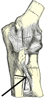
The oblique cord is a ligament between the ulnar and radius bones in the lower arm near the elbow. It takes the form of a small, flattened band, extending downward and lateralward, from the lateral side of the ulnar tuberosity at the base of the coronoid process to the radius a little below the radial tuberosity. Its fibers run in the opposite direction to those of the Interosseous membrane of the forearm.
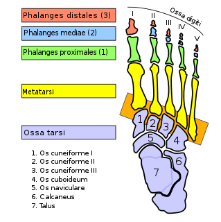
The tarsometatarsal joints are arthrodial joints in the foot. The tarsometatarsal joints involve the first, second and third cuneiform bones, the cuboid bone and the metatarsal bones. The eponym Lisfranc joint is named after 18th-19th century surgeon and gynecologist, Jacques Lisfranc de St. Martin.

The anterior interosseous nerve is a branch of the median nerve that supplies the deep muscles on the anterior of the forearm, except the ulnar (medial) half of the flexor digitorum profundus. Its nerve roots come from C8 and T1.
The interosseous membrane of the forearm is a fibrous sheet that connects the interosseous margins of the radius and the ulna. It is the main part of the radio-ulnar syndesmosis, a fibrous joint between the two bones.
The posterior compartment of the forearm contains twelve muscles which are chiefly responsible for extension of the wrist and digits, and supination of the forearm. It is separated from the anterior compartment by the interosseous membrane between the radius and ulna.

In human anatomy, the radial (RCL) and ulnar (UCL) collateral ligaments of the metacarpophalangeal joints (MCP) of the hand are the primary stabilisers of the MCP joints. They have two parts: the cord-like collateral ligaments proper located more dorsally and the accessory collateral ligaments located more volarly. They enable us to spread our fingers with an open hand but not with the hand closed into a fist.
This page is based on this
Wikipedia article Text is available under the
CC BY-SA 4.0 license; additional terms may apply.
Images, videos and audio are available under their respective licenses.