Subclavius muscle

Last updated
Subclavius muscle
Subclavius muscle frontal2.png
Subclavius muscle (shown in red).
Gray411subclavius.png
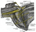
Deep muscles of the chest and front of the arm, with the boundaries of the axilla. (Subclavius visible at upper left, above first rib.)
Details
Origin first rib and cartilage
Insertion subclavian groove of clavicle (inferior surface of middle one third of the clavicle)
Artery thoracoacromial trunk, clavicular branch
Nerve subclavian nerve
Actions depression of clavicle
elevation of first rib
Identifiers
Latin musculus subclavius
TA98 A04.4.01.007
TA2 2306
FMA 13410
Anatomical terms of muscle

The subclavius is a small triangular muscle, placed between the clavicle and the first rib. [1] Along with the pectoralis major and pectoralis minor muscles, the subclavius muscle makes up the anterior axioappendicular muscles, also known as anterior wall of the axilla. [2]

Contents

Structure

It arises by a short, thick tendon from the first rib and its cartilage at their junction, in front of the costoclavicular ligament. [1]

The fleshy fibers proceed obliquely superolaterally, to be inserted into the groove on the under surface of the clavicle.

Innervation

The nerve to subclavius (or subclavian nerve) innervates the muscle. This arises from the junction of the fifth and sixth cervical nerves, from the superior/upper trunk of the brachial plexus.

Variation

Insertion into coracoid process instead of clavicle or into both clavicle and coracoid process. Sternoscapular fasciculus to the upper border of scapula. Sternoclavicularis [3] from manubrium to clavicle between pectoralis major and coracoclavicular fascia. [1] Rarely, the subclavius may be missing entirely. [4]

Function

It depresses the lateral clavicle, acts to stabilize the clavicle while the shoulder moves the arm. It also raises the first rib while lowering the clavicle during breathing.

The subclavius protects the underlying brachial plexus and subclavian vessels from a broken clavicle - the most frequently broken long bone [5]

Additional images

Related Research Articles

Arm Proximal part of the free upper limb between the shoulder and the elbow

In human anatomy, the arm is the part of the upper limb between the glenohumeral joint and the elbow joint. In common usage, the arm extends through the hand. The arm can be divided into the upper arm, which extends from the shoulder to the elbow, the forearm which extends from the elbow to the hand, and the hand. Anatomically the shoulder girdle with bones and corresponding muscles is by definition a part of the arm. The Latin term brachium may refer to either the arm as a whole or to the upper arm on its own.

Scapula Bone that connects the humerus (upper arm bone) with the clavicle (collar bone)

In anatomy, the scapula, also known as the shoulder bone, shoulder blade, wing bone, speal bone or blade bone, is the bone that connects the humerus with the clavicle. Like their connected bones, the scapulae are paired, with each scapula on either side of the body being roughly a mirror image of the other. The name derives from the Classical Latin word for trowel or small shovel, which it was thought to resemble.

Brachial plexus

The brachial plexus is a network of nerves (formed by the anterior rami of the lower four cervical nerves and first thoracic nerve. This plexus extends from the spinal cord, through the cervicoaxillary canal in the neck, over the first rib, and into the armpit. It supplies afferent and efferent nerve fibers to the chest, shoulder, arm, forearm, and hand.

Coracoid process Small hook-like structure on the lateral edge of the superior anterior portion of the scapula

The coracoid process is a small hook-like structure on the lateral edge of the superior anterior portion of the scapula. Pointing laterally forward, it, together with the acromion, serves to stabilize the shoulder joint. It is palpable in the deltopectoral groove between the deltoid and pectoralis major muscles.

Subclavian artery Major arteries of the upper thorax, below the clavicle

In human anatomy, the subclavian arteries are paired major arteries of the upper thorax, below the clavicle. They receive blood from the aortic arch. The left subclavian artery supplies blood to the left arm and the right subclavian artery supplies blood to the right arm, with some branches supplying the head and thorax. On the left side of the body, the subclavian comes directly off the aortic arch, while on the right side it arises from the relatively short brachiocephalic artery when it bifurcates into the subclavian and the right common carotid artery.

Levator scapulae muscle

The levator scapulae is a skeletal muscle situated at the back and side of the neck. As the Latin name suggests, its main function is to lift the scapula.

Pectoralis major

The pectoralis major is a thick, fan-shaped or triangular convergent muscle, situated at the chest of the human body. It makes up the bulk of the chest muscles and lies under the breast. Beneath the pectoralis major is the pectoralis minor, a thin, triangular muscle. The pectoralis major's primary functions are flexion, adduction, and internal rotation of the humerus. The pectoral major may colloquially be referred to as "pecs", "pectoral muscle" or "chest muscle" due to it being the largest and most superficial muscle in the chest area.

Pectoralis minor human chest muscle that protracts the shoulder

Pectoralis minor muscle is a thin, triangular muscle, situated at the upper part of the chest, beneath the pectoralis major in the human body.

Suprascapular nerve

The suprascapular nerve is a nerve that branches from the upper trunk of the brachial plexus. It is responsible for the innervation of two of the muscles that originate from the scapula, namely the supraspinatus and infraspinatus muscles.

Serratus anterior muscle Muscle on the surface of the ribs

The serratus anterior is a muscle that originates on the surface of the 1st to 8th ribs at the side of the chest and inserts along the entire anterior length of the medial border of the scapula. The serratus anterior acts to pull the scapula forward around the thorax. The muscle is named from Latin: serrare = to saw, referring to the shape, anterior = on the front side of the body.

Axillary artery Large blood vessel bringing oxygenated blood to the thorax

In human anatomy, the axillary artery is a large blood vessel that conveys oxygenated blood to the lateral aspect of the thorax, the axilla (armpit) and the upper limb. Its origin is at the lateral margin of the first rib, before which it is called the subclavian artery.

Scalene muscles Muscles on the sides of the neck

The scalene muscles are a group of three pairs of muscles in the lateral neck, namely the anterior scalene, middle scalene, and posterior scalene. They are innervated by the fourth, fifth, and sixth cervical spinal nerves (C4-C6).

Shoulder joint Synovial ball and socket joint in the shoulder

The shoulder joint is structurally classified as a synovial ball-and-socket joint and functionally as a diarthrosis and multiaxial joint. It involves an articulation between the glenoid fossa of the scapula and the head of the humerus. Due to the very loose joint capsule that gives a limited interface of the humerus and scapula, it is the most mobile joint of the human body.

Coracobrachialis muscle

The coracobrachialis muscle is the smallest of the three muscles that attach to the coracoid process of the scapula. It is situated at the upper and medial part of the arm.

Medial pectoral nerve

The medial pectoral nerve arises from the medial cord of the brachial plexus, and through it from the eighth cervical and first thoracic roots (C8/T1).

Suprascapular artery Artery of the neck

The suprascapular artery is a branch of the thyrocervical trunk on the neck.

Clavipectoral fascia

The clavipectoral fascia is a strong fascia situated under cover of the clavicular portion of the pectoralis major.

Subclavian nerve

The subclavian nerve, also known as the nerve to the subclavius, is small branch of the upper trunk of the brachial plexus. It contains axons from C5 and C6. The subclavian nerve innervates the subclavius muscle.

Subclavian triangle

The subclavian triangle, the smaller division of the posterior triangle, is bounded, above, by the inferior belly of the omohyoideus; below, by the clavicle; its base is formed by the posterior border of the sternocleidomastoideus.

Clavipectoral triangle

The clavipectoral triangle is an anatomical region found in humans and other animals. It is bordered by the following structures:

References

PD-icon.svgThis article incorporates text in the public domain from page 438 of the 20th edition of Gray's Anatomy (1918)

  1. 1 2 3 "IV. Myology: 13". Gray's Anatomy: The Muscles Connecting the Upper Extremity to the Anterior and Lateral Thoracic Walls. 1918. Archived from the original on 2014-03-08.
  2. Drake, Richard, et al. Gray's Anatomy For Students, Elsevier Inc., 2005
  3. Sternoclavicularis is a rare muscle found in a large triangular gap between the sternocostal and clavicularheads of Pectoralis Major muscle on the right side during routine cadaveric dissection.
  4. Yun, Sam; Park, Sekyoung; Kim, Chang Su (May 2018). "Absence of the subclavius muscle with contralateral subclavius posticus muscle: first imaging report". Clinical Imaging. 49: 54–57. doi:10.1016/j.clinimag.2017.10.008. ISSN   0899-7071. PMID   29127878.
  5. ?