External intercostal muscles

Last updated
External intercostal muscles
External intercostal muscles back.png
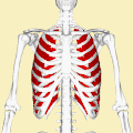
Position of the external intercostal muscles (shown in red) seen from the back.
Details
Origin Lower border of ribs
Insertion Upper border of rib below
Artery Intercostal arteries
Nerve Intercostal nerves
Actions Inhalation
Antagonist Intercostales interni muscles
Identifiers
Latin musculi intercostales externi
TA98 A04.4.01.012
TA2 2311
FMA 74084
Anatomical terms of muscle

The external intercostal muscles or external intercostals (intercostales externi) are eleven in number on both sides.

Contents

Structure

A cutout of the thoracic wall showing the three layers of intercostal muscle - from the left wall. 1114 Thorax zoom.png
A cutout of the thoracic wall showing the three layers of intercostal muscle - from the left wall.

The muscles extend from the tubercles of the ribs behind, to the cartilages of the ribs in front, where they end in thin membranes, the external intercostal membranes, which are continued forward to the sternum. These muscles work in unison when inhalation occurs. The internal intercostal muscles relax while the external muscles contract causing the expansion of the chest cavity and an influx of air into the lungs.

Each arises from the lower border of a rib, and is inserted into the upper border of the rib below. In the two lower spaces they extend to the ends of the cartilages, and in the upper two or three spaces they do not quite reach the ends of the ribs.

They are thicker than the internal intercostals, and their fibers are directed obliquely downward and laterally on the back of the thorax, and downward, forward, and medially on the front.

Variations

Continuation with the external oblique or serratus anterior: A supracostalis muscle, from the anterior end of the first rib down to the second, third or fourth ribs occasionally occurs.

Additional images

See also

Related Research Articles

<span class="mw-page-title-main">Rib cage</span> Bone structure of the thorax

The rib cage or thoracic cage is an endoskeletal enclosure in the thorax of most vertebrates that comprises the ribs, vertebral column and sternum, which protect the vital organs of the thoracic cavity, such as the heart, lungs and great vessels and support the shoulder girdle to form the core part of the axial skeleton.

<span class="mw-page-title-main">Humerus</span> Long bone of the upper arm

The humerus is a long bone in the arm that runs from the shoulder to the elbow. It connects the scapula and the two bones of the lower arm, the radius and ulna, and consists of three sections. The humeral upper extremity consists of a rounded head, a narrow neck, and two short processes. The body is cylindrical in its upper portion, and more prismatic below. The lower extremity consists of 2 epicondyles, 2 processes, and 3 fossae. As well as its true anatomical neck, the constriction below the greater and lesser tubercles of the humerus is referred to as its surgical neck due to its tendency to fracture, thus often becoming the focus of surgeons.

<span class="mw-page-title-main">Thorax</span> Frontal part of an animals body, between its head and abdomen

The thorax or chest is a part of the anatomy of mammals and other tetrapod animals located between the neck and the abdomen. In insects, crustaceans, and the extinct trilobites, the thorax is one of the three main divisions of the creature's body, each of which is in turn composed of multiple segments.

<span class="mw-page-title-main">Intercostal muscles</span> Muscle groups between the ribs which form and move the chest wall during breathing

The intercostal muscles comprise many different groups of muscles that run between the ribs, and help form and move the chest wall. The intercostal muscles are mainly involved in the mechanical aspect of breathing by helping expand and shrink the size of the chest cavity.

<span class="mw-page-title-main">Pectoralis major</span> Main human chest muscle

The pectoralis major is a thick, fan-shaped or triangular convergent muscle of the human chest. It makes up the bulk of the chest muscles and lies under the breast. Beneath the pectoralis major is the pectoralis minor muscle.

<span class="mw-page-title-main">Rectus abdominis muscle</span> Paired straight muscle

The rectus abdominis muscle, also known as the "abdominal muscle" or simply the "abs", is a pair of segmented skeletal muscle on the ventral aspect of a person's abdomen. The paired muscle is separated at the midline by a band of dense connective tissue called the linea alba, and the connective tissue defining each lateral margin of the rectus abdominus is the linea semilunaris. The muscle extends from the pubic symphysis, pubic crest and pubic tubercle inferiorly, to the xiphoid process and costal cartilages of the 5th–7th ribs superiorly.

<span class="mw-page-title-main">Transverse abdominal muscle</span> Muscle of the abdominal area

The transverse abdominal muscle (TVA), also known as the transverse abdominis, transversalis muscle and transversus abdominis muscle, is a muscle layer of the anterior and lateral abdominal wall, deep to the internal oblique muscle. It is thought by most fitness instructors to be a significant component of the core.

<span class="mw-page-title-main">Transversus thoracis muscle</span>

The transversus thoracis muscle, also known as triangularis sterni, lies internal to the thoracic cage, anteriorly. It is usually a thin plane of muscular and tendinous fibers, however on athletic individuals it can be a thick 'slab of meat', situated upon the inner surface of the front wall of the chest. It is in the same layer as the subcostal muscles and the innermost intercostal muscles.

<span class="mw-page-title-main">Abdominal internal oblique muscle</span> Muscle in the abdominal wall

The abdominal internal oblique muscle, also internal oblique muscle or interior oblique, is an abdominal muscle in the abdominal wall that lies below the external oblique muscle and just above the transverse abdominal muscle.

<span class="mw-page-title-main">Internal intercostal muscles</span> Group of muscles between the ribs

The internal intercostal muscles are a group of skeletal muscles located between the ribs. They are eleven in number on either side. They commence anteriorly at the sternum, in the intercostal spaces between the cartilages of the true ribs, and at the anterior extremities of the cartilages of the false ribs, and extend backward as far as the angles of the ribs, hence they are continued to the vertebral column by thin aponeuroses, the posterior intercostal membranes. They pull the sternum and ribs upward and inward.

<span class="mw-page-title-main">Abdominal external oblique muscle</span> Skeletal muscle in the abdomen

The abdominal external oblique muscle is the largest and outermost of the three flat abdominal muscles of the lateral anterior abdomen.

<span class="mw-page-title-main">Intercostal nerves</span> Nerves in the thorax and abdomen

The intercostal nerves are part of the somatic nervous system, and arise from the anterior rami of the thoracic spinal nerves from T1 to T11. The intercostal nerves are distributed chiefly to the thoracic pleura and abdominal peritoneum, and differ from the anterior rami of the other spinal nerves in that each pursues an independent course without plexus formation.

<span class="mw-page-title-main">Abdomen</span> Part of the body between the chest and pelvis

The abdomen is the part of the body between the thorax (chest) and pelvis, in humans and in other vertebrates. The abdomen is the front part of the abdominal segment of the torso. The area occupied by the abdomen is called the abdominal cavity. In arthropods, it is the posterior tagma of the body; it follows the thorax or cephalothorax.

<span class="mw-page-title-main">Pubis (bone)</span> Most forward-facing of the three main regions making up the os coxa

In vertebrates, the pubis or pubic bone forms the lower and anterior part of each side of the hip bone. The pubis is the most forward-facing of the three bones that make up the hip bone. The left and right pubic bones are each made up of three sections; A superior ramus, inferior ramus, and a body.

<span class="mw-page-title-main">Costal cartilage</span> Resilient, smooth, glass-like tissue at the front ends of ribs in verterbrates

The costal cartilages are bars of hyaline cartilage that serve to prolong the ribs forward and contribute to the elasticity of the walls of the thorax. Costal cartilage is only found at the anterior ends of the ribs, providing medial extension.

<span class="mw-page-title-main">Intercostal space</span> Anatomic space between two ribs

The intercostal space (ICS) is the anatomic space between two ribs. Since there are 12 ribs on each side, there are 11 intercostal spaces, each numbered for the rib superior to it.

Unlike the other two intercostal muscles, the external intercostal muscle does not retain its muscular character all the way to the sternum, and so the tissue in this location is called the external intercostal membrane.

<span class="mw-page-title-main">Muscles of respiration</span> Muscles involved in breathing

The muscles of respiration are the muscles that contribute to inhalation and exhalation, by aiding in the expansion and contraction of the thoracic cavity. The diaphragm and, to a lesser extent, the intercostal muscles drive respiration during quiet breathing. The elasticity of these muscles is crucial to the health of the respiratory system and to maximize its functional capabilities.

<span class="mw-page-title-main">Sternum</span> Flat bone in the middle front part of the rib cage

The sternum or breastbone is a long flat bone located in the central part of the chest. It connects to the ribs via cartilage and forms the front of the rib cage, thus helping to protect the heart, lungs, and major blood vessels from injury. Shaped roughly like a necktie, it is one of the largest and longest flat bones of the body. Its three regions are the manubrium, the body, and the xiphoid process. The word sternum originates from Ancient Greek στέρνον (stérnon) 'chest'.

<span class="mw-page-title-main">Pulmonary pleurae</span> Membrane lining the thoracic cavity wall

The pulmonary pleurae are the two flattened sacs ensheathing each lung, locally appearing as two opposing layers of serous membrane separating the lungs from the mediastinum and the inside surfaces of the surrounding chest walls.

References

PD-icon.svgThis article incorporates text in the public domain from page 403 of the 20th edition of Gray's Anatomy (1918)