Sternalis muscle

Last updated
Sternalis
Grant 1962 27b.png
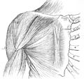
Sternalis muscle, in line with rectus abdominis and sternomastoid, was found in 6% of 535 cadavera (R. N. Barlow)
Details
Origin Manubrium of sternum or clavicle
Insertion Xiphoid process, pectoral fascia, lower ribs, costal cartilages or rectus sheath
Identifiers
Latin musculus sternalis
TA98 A04.4.01.001
TA2 2300
FMA 9717
Anatomical terms of muscle

The sternalismuscle is an anatomical variation that lies in front of the sternal end of the pectoralis major parallel to the margin of the sternum. The sternalis muscle may be a variation of the pectoralis major or of the rectus abdominis.

Contents

Structure

The sternalis is a muscle that runs along the anterior aspect of the body of the sternum. It lies superficially and parallel to the sternum. Its origin and insertion are variable. The sternalis muscle often originates from the upper part of the sternum and can display varying insertions such as the pectoral fascia, lower ribs, costal cartilages, rectus sheath, aponeurosis of the abdominal external oblique muscle. [1] [2] [3] There is still a great deal of disagreement about its innervation and its embryonic origin. [1]

In a review, [4] it was reported that the muscle was innervated by the external or internal thoracic nerves in 55% of the cases, by the intercostal nerves in 43% of the cases, while the remaining cases were supplied by both nerves. [4] However, innervation by the pectoral nerves has also been reported. [5] This appears to indicate that the sternalis is not always derived from the same embryonic origin. [6]

Prevalence

Cadaveric studies showed that the sternalis muscle has a mean prevalence of around 7.8% in the population [7] with the range from 0.5% to 23.5%. [8] It has a slightly higher incidence in females. [1] Though, it was proposed that a possible reason for the high prevalence may result from the existence of small, ill-defined or tendinous fibres, which could be misidentified for a sternalis muscle. [9]

Variations

A 2014 study [8] classified the sternalis into three types depending on morphology.

Type I, the single head and single belly was seen in the majority of reported cases (58.5%), type II in 18.1%, and type III in 23.4%. [8]

In addition to the above classification, triple-bellied/double-headed sternalis has also been reported. [10]

Function

There is no apparent physiological function of the sternalis muscle. [11] However, there are many theories for a function. It may function as a proprioceptive sensor for thoracic wall movements. [12] It may also take part in the movement of the shoulder joint or have an additional role in elevation of the chest wall. [13]

Clinical significance

The presence of the sternalis in sagittal plane on CT (left) and on volume rendered CT 3D reconstruction (right). Musculus sternalis rechts - CT sagittal und Volume rendering - 001.jpg
The presence of the sternalis in sagittal plane on CT (left) and on volume rendered CT 3D reconstruction (right).

The presence of the sternalis is asymptomatic [1] but aesthetic complaints have been reported as it was reported to cause chest asymmetry or deviation of the nipple-areola complex. [1] [14] The presence of the sternalis may cause alterations in the electrocardiogram [15] or confusion in mammography. [16] However, there is a potential benefit of the muscle as it can be used as a flap in a reconstructive surgery of the head and neck and the anterior chest wall. [14]

History

The sternalis was first reported by Carbolius in 1604 and the name was first given by Turner in 1867. [17] Different terminologies have been given to the sternalis due to its highly varied morphology and the disagreement on its embryonic origin. The sternalis was referred to as the rectus sternalis,sternalis brutorum, musculus sternalis, episternalis, parasternalis, presternalis, rectus sterni, rectus thoracis, rectus thoracicus superficialis, superficial rectus abdominis, japonicas, and thoracicus depending on studies. [7] [17]

Additional images

Related Research Articles

<span class="mw-page-title-main">Abducens nerve</span> Cranial nerve VI, for eye movements

The abducens nerve or abducent nerve, also known as the sixth cranial nerve, cranial nerve VI, or simply CN VI, is a cranial nerve in humans and various other animals that controls the movement of the lateral rectus muscle, one of the extraocular muscles responsible for outward gaze. It is a somatic efferent nerve.

<span class="mw-page-title-main">Radial nerve</span> Nerve in the human body that supplies the posterior portion of the upper limb

The radial nerve is a nerve in the human body that supplies the posterior portion of the upper limb. It innervates the medial and lateral heads of the triceps brachii muscle of the arm, as well as all 12 muscles in the posterior osteofascial compartment of the forearm and the associated joints and overlying skin.

<span class="mw-page-title-main">Pectoralis major</span> Main human chest muscle

The pectoralis major is a thick, fan-shaped or triangular convergent muscle of the human chest. It makes up the bulk of the chest muscles and lies under the breast. Beneath the pectoralis major is the pectoralis minor muscle.

<span class="mw-page-title-main">Aponeurosis</span> Tissue which connects muscles to other organs

An aponeurosis is a flattened tendon by which muscle attaches to bone or fascia. Aponeuroses exhibit an ordered arrangement of collagen fibres, thus attaining high tensile strength in a particular direction while being vulnerable to tensional or shear forces in other directions. They have a shiny, whitish-silvery color, are histologically similar to tendons, and are very sparingly supplied with blood vessels and nerves. When dissected, aponeuroses are papery and peel off by sections. The primary regions with thick aponeuroses are in the ventral abdominal region, the dorsal lumbar region, the ventriculus in birds, and the palmar (palms) and plantar (soles) regions.

<span class="mw-page-title-main">Rectus abdominis muscle</span> Paired straight muscle

The rectus abdominis muscle, also known as the "abdominal muscle" or simply the "abs", is a pair of segmented skeletal muscle on the ventral aspect of a person's abdomen. The paired muscle is separated at the midline by a band of dense connective tissue called the linea alba, and the connective tissue defining each lateral margin of the rectus abdominus is the linea semilunaris. The muscle extends from the pubic symphysis, pubic crest and pubic tubercle inferiorly, to the xiphoid process and costal cartilages of the 5th–7th ribs superiorly.

<span class="mw-page-title-main">Superior rectus muscle</span> Extraocular muscle that elevates the eye

The superior rectus muscle is a muscle in the orbit. It is one of the extraocular muscles. It is innervated by the superior division of the oculomotor nerve (III). In the primary position, its primary function is elevation, although it also contributes to intorsion and adduction. It is associated with a number of medical conditions, and may be weak, paralysed, overreactive, or even congenitally absent in some people.

<span class="mw-page-title-main">Medial rectus muscle</span> Extraocular muscle that rotates the eye medially

The medial rectus muscle is a muscle in the orbit near the eye. It is one of the extraocular muscles. It originates from the common tendinous ring, and inserts into the anteromedial surface of the eye. It is supplied by the inferior division of the oculomotor nerve (III). It rotates the eye medially (adduction).

<span class="mw-page-title-main">Transverse abdominal muscle</span> Muscle of the abdominal area

The transverse abdominal muscle (TVA), also known as the transverse abdominis, transversalis muscle and transversus abdominis muscle, is a muscle layer of the anterior and lateral abdominal wall, deep to the internal oblique muscle. It is thought by most fitness instructors to be a significant component of the core.

<span class="mw-page-title-main">Intermammary cleft</span> Breast cleavage

The intermammary cleft, intermammary sulcus, or sulcus intermammarius is a surface feature of males and females that marks the division of the two breasts with the sternum (breastbone) in the middle. The International Federation of Associations of Anatomists (IFAA) uses the terms "sulcus intermammarius" or "intermammary cleft" when referring to the area between the breasts.

<span class="mw-page-title-main">Subclavius muscle</span> Muscle between the clavicle and first rib

The subclavius is a small triangular muscle, placed between the clavicle and the first rib. Along with the pectoralis major and pectoralis minor muscles, the subclavius muscle makes up the anterior axioappendicular muscles, also known as anterior wall of the axilla.

<span class="mw-page-title-main">Popliteus muscle</span> Muscle responsible for unlocking the knees during walking

The popliteus muscle in the leg is used for unlocking the knees when walking, by laterally rotating the femur on the tibia during the closed chain portion of the gait cycle. In open chain movements, the popliteus muscle medially rotates the tibia on the femur. It is also used when sitting down and standing up. It is the only muscle in the posterior (back) compartment of the lower leg that acts just on the knee and not on the ankle. The gastrocnemius muscle acts on both joints.

<span class="mw-page-title-main">Lateral pectoral nerve</span>

The lateral pectoral nerve arises from the lateral cord of the brachial plexus, and through it from the C5-7.

<span class="mw-page-title-main">Pectoral fascia</span>

The pectoral fascia is a thin lamina, covering the surface of the pectoralis major, and sending numerous prolongations between its fasciculi: it is attached, in the middle line, to the front of the sternum; above, to the clavicle; laterally and below it is continuous with the fascia of the shoulder, axilla, and thorax.

<span class="mw-page-title-main">Pectineal ligament</span>

The pectineal ligament, sometimes known as the inguinal ligament of Cooper, is an extension of the lacunar ligament. It runs on the pectineal line of the pubic bone. The pectineal ligament is the posterior border of the femoral ring.

The linea semilunaris is described by Adriaan van den Spiegel described the Linea Semilunaris as the line forming and marking the transition from muscle to aponeurosis in the transversus abdominis muscle of the abdomen. It needs to be distinguished from the lateral border of the rectus abdominis muscle sheath which is often wrongly described as semilunar line. The lateral border of the rectus sheath is part of the recently described EIT ambivium to more precisely describe this important part of the anterior abdominal wall. The EIT ambivium is formed by the Musculus Obliquus Externus (MOE), the Musculus Obliquus Internus (MOI) and the Musculus Transversus Abdominis (TA). Understanding the interrelationship between the lateral muscles and the medial compartment is of paramount importance for surgeons to understand the surgical anatomy.

An accessory muscle is a relatively rare anatomical variation where duplication of a muscle may appear anywhere in the muscular system. Treatment is not indicated unless the accessory muscle interferes with normal function.

<span class="mw-page-title-main">Extensor indicis et medii communis muscle</span>

The extensor indicis et medii communis is a rare anatomical variant in the extensor compartment of forearm. This additional muscle lies in the deep extensor layer next to the extensor indicis proprius and the extensor pollicis longus. The characteristics of this anomalous muscle resemble those of the extensor indicis proprius, with split tendons to the index and the middle finger. This muscle can also be considered as a variation of the aberrant extensor medii proprius.

<span class="mw-page-title-main">Axillary arch</span> Muscular slip associated with latissimus dorsi muscle

The axillary arch is a variant of the latissimus dorsi muscle in humans. It is found as a slip of muscle or fascia extending between the latissimus dorsi muscle and the pectoralis major. There is considerable variation in the exact position of its origin and insertions as well as its blood and nerve supply. The arch may occur on one or both sides of the body. A meta-analysis revealed that the axillary arch had an overall prevalence of 5.3% of limbs.

An anatomical variation, anatomical variant, or anatomical variability is a presentation of body structure with morphological features different from those that are typically described in the majority of individuals. Anatomical variations are categorized into three types including morphometric, consistency, and spatial.

The myodural bridge or miodural ligament is a bridge of connective tissue that extends between the suboccipital muscles and the cervical spinal dura mater, the outer membrane that envelops the spinal cord. It provides a physical connection between the musculoskeletal and nervous systems, and the circulation of cerebrospinal fluid. Its importance has been highlighted by various authors.

References

  1. 1 2 3 4 5 Raikos, Athanasios; Paraskevas, George K.; Yusuf, Faisal; Kordali, Panagiota; Ioannidis, Orestis; Brand-Saberi, Beate (2011-12-01). "Sternalis muscle: a new crossed subtype, classification, and surgical applications". Annals of Plastic Surgery. 67 (6): 646–648. doi:10.1097/SAP.0b013e31820d688b. ISSN   1536-3708. PMID   21407048.
  2. Georgiev, Georgi P.; Jelev, Lazar; Ovtscharoff, Vladimir A. (2009-09-01). "On the clinical significance of the sternalis muscle". Folia Medica. 51 (3): 53–56. ISSN   0204-8043. PMID   19957564.
  3. "Anatomy 2008; 2: 67-71 [Case Report]". www.anatomy.org.tr. Retrieved 2015-09-05.
  4. 1 2 O'NEILL, M. N.; FOLAN-CURRAN, J. (1998-08-01). "Case report: bilateral sternalis muscles with a bilateral pectoralis major anomaly". Journal of Anatomy. 193 (Pt 2): 289–292. doi:10.1046/j.1469-7580.1998.19320289.x. ISSN   0021-8782. PMC   1467849 . PMID   9827645.
  5. Kida, M. Y.; Izumi, A.; Tanaka, S. (2000-01-01). "Sternalis muscle: topic for debate". Clinical Anatomy. 13 (2): 138–140. doi:10.1002/(SICI)1098-2353(2000)13:2<138::AID-CA8>3.0.CO;2-4. ISSN   0897-3806. PMID   10679858.
  6. Harper WF. The sternalis muscle in the anencephalous foetus. Anat Notes 1936;317-20.
  7. 1 2 Snosek, Michael; Tubbs, R. Shane; Loukas, Marios (2014-09-01). "Sternalis muscle, what every anatomist and clinician should know". Clinical Anatomy. 27 (6): 866–884. doi:10.1002/ca.22361. ISSN   1098-2353. PMID   24431029.
  8. 1 2 3 Ge, Zufeng; Tong, Yunlong; Zhu, Shiqiang; Fang, Xiong; Zhuo, Lang; Gong, Xiangyang (2014-04-01). "Prevalence and variance of the sternalis muscle: a study in the Chinese population using multi-detector CT". Surgical and Radiologic Anatomy. 36 (3): 219–224. doi:10.1007/s00276-013-1175-4. ISSN   1279-8517. PMID   23912561.
  9. JELEV, L.; GEORGIEV, G.; SURCHEV, L. (2001-09-01). "The sternalis muscle in the Bulgarian population: classification of sternales". Journal of Anatomy. 199 (Pt 3): 359–363. doi:10.1046/j.1469-7580.2001.19930359.x. ISSN   0021-8782. PMC   1468341 . PMID   11554516.
  10. Dudgeon, Sarah N.; Marcotte, Kayla M.; Fox, Glenn M.; Alsup, B. Kathleen (2017-12-01). "A previously unclassified variant of sternalis muscle". Surgical and Radiologic Anatomy. 39 (12): 1417–1419. doi:10.1007/s00276-017-1883-2. ISSN   0930-1038. PMID   28593340.
  11. Snosek, M; Tubbs, RS; Loukas, M (September 2014). "Sternalis muscle, what every anatomist and clinician should know". Clinical Anatomy. 27 (6): 866–84. doi:10.1002/ca.22361. PMID   24431029.
  12. Natsis, Konstantinos; Totlis, Trifon (2007-11-01). "A rare accessory muscle of the anterior thoracic wall". Clinical Anatomy. 20 (8): 980–981. doi:10.1002/ca.20534. ISSN   0897-3806. PMID   17948292.
  13. Young Lee, Bae; Young Byun, Jae; Hee Kim, Hak; Sook Kim, Hyun; Mee Cho, Song; Hoon Lee, Kang; Sup Song, Kyung; Soo Kim, Bum; Mun Lee, Jae (2006-08-01). "The sternalis muscles: incidence and imaging findings on MDCT". Journal of Thoracic Imaging. 21 (3): 179–183. doi:10.1097/01.rti.0000208287.04490.db. ISSN   0883-5993. PMID   16915061.
  14. 1 2 Bailey, P. M.; Tzarnas, C. D. (1999-04-01). "The sternalis muscle: a normal finding encountered during breast surgery". Plastic and Reconstructive Surgery. 103 (4): 1189–1190. doi:10.1097/00006534-199904040-00013. ISSN   0032-1052. PMID   10088505.
  15. Arráez-Aybar, L. A.; Sobrado-Perez, J.; Merida-Velasco, J. R. (2003-07-01). "Left musculus sternalis". Clinical Anatomy. 16 (4): 350–354. doi:10.1002/ca.10120. ISSN   0897-3806. PMID   12794922.
  16. Bradley, F. M.; Hoover, H. C.; Hulka, C. A.; Whitman, G. J.; McCarthy, K. A.; Hall, D. A.; Moore, R.; Kopans, D. B. (1996-01-01). "The sternalis muscle: an unusual normal finding seen on mammography". AJR. American Journal of Roentgenology. 166 (1): 33–36. doi:10.2214/ajr.166.1.8571900. ISSN   0361-803X. PMID   8571900.
  17. 1 2 Turner W (1867) On the musculus sternalis. J Anat Physiol 1(2):246–378