
In vertebrate anatomy, ribs are the long curved bones which form the rib cage, part of the axial skeleton. In most tetrapods, ribs surround the chest, enabling the lungs to expand and thus facilitate breathing by expanding the chest cavity. They serve to protect the lungs, heart, and other internal organs of the thorax. In some animals, especially snakes, ribs may provide support and protection for the entire body.
The rib cage is an endoskeletal enclosure in the thorax of most vertebrate animals that comprises the ribs, vertebral column and sternum, which protects vital organs such as the heart, lungs and great vessels. The circumferential enclosure formed by left and right rib cages, together known as the thoracic cage, is a semi-rigid bony and cartilaginous structure which surrounds the thoracic cavity and supports the shoulder girdles to form the core part of the axial skeleton.

The trapezius is a large paired trapezoid-shaped surface muscle that extends longitudinally from the occipital bone to the lower thoracic vertebrae of the spine and laterally to the spine of the scapula. It moves the scapula and supports the arm.

A spinal nerve is a mixed nerve, which carries motor, sensory, and autonomic signals between the spinal cord and the body. In the human body there are 31 pairs of spinal nerves, one on each side of the vertebral column. These are grouped into the corresponding cervical, thoracic, lumbar, sacral and coccygeal regions of the spine. There are eight pairs of cervical nerves, twelve pairs of thoracic nerves, five pairs of lumbar nerves, five pairs of sacral nerves, and one pair of coccygeal nerves. The spinal nerves are part of the peripheral nervous system.

The lumbar vertebrae are, in human anatomy, the five vertebrae between the rib cage and the pelvis. They are the largest segments of the vertebral column and are characterized by the absence of the foramen transversarium within the transverse process and by the absence of facets on the sides of the body. They are designated L1 to L5, starting at the top. The lumbar vertebrae help support the weight of the body, and permit movement.

The thoracic diaphragm, or simply the diaphragm, is a sheet of internal skeletal muscle in humans and other mammals that extends across the bottom of the thoracic cavity. The diaphragm is the most important muscle of respiration, and separates the thoracic cavity, containing the heart and lungs, from the abdominal cavity: as the diaphragm contracts, the volume of the thoracic cavity increases, creating a negative pressure there, which draws air into the lungs. Its high oxygen consumption is noted by the many mitochondria and capillaries present; more than in any other skeletal muscle.
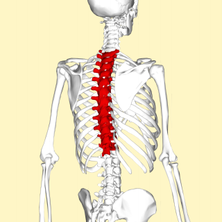
In vertebrates, thoracic vertebrae compose the middle segment of the vertebral column, between the cervical vertebrae and the lumbar vertebrae. In humans, there are twelve thoracic vertebrae and they are intermediate in size between the cervical and lumbar vertebrae; they increase in size going towards the lumbar vertebrae, with the lower ones being much larger than the upper. They are distinguished by the presence of facets on the sides of the bodies for articulation with the heads of the ribs, as well as facets on the transverse processes of all, except the eleventh and twelfth, for articulation with the tubercles of the ribs. By convention, the human thoracic vertebrae are numbered T1–T12, with the first one (T1) located closest to the skull and the others going down the spine toward the lumbar region.
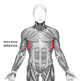
The serratus anterior is a muscle of the chest. It originates at the side of the chest from the upper 8 or 9 ribs; it inserts along the entire length of the anterior aspect of the medial border of the scapula. It is innervated by the long thoracic nerve from the brachial plexus. The serratus anterior acts to pull the scapula forward around the thorax.

The scalene muscles are a group of three muscles on each side of the neck, identified as the anterior, the middle, and the posterior. They are innervated by the third to the eighth cervical spinal nerves (C3-C8).

The multifidusmuscle consists of a number of fleshy and tendinous fasciculi, which fill up the groove on either side of the spinous processes of the vertebrae, from the sacrum to the axis. While very thin, the multifidus muscle plays an important role in stabilizing the joints within the spine. The multifidus is one of the transversospinales.

The serratus posterior superior muscle is a thin, quadrilateral muscle. It is situated at the upper back part of the thorax, deep to the rhomboid muscles.

The abdominal external oblique muscle is the largest and outermost of the three flat abdominal muscles of the lateral anterior abdomen.

The erector spinae or spinal erectors is a set of muscles that straighten and rotate the back. The spinal erectors work together with the glutes to maintain stable posture standing or sitting.

The thoracolumbar fascia is a complex, multilayer arrangement of fascial and aponeurotic layers forming a separation between the paraspinal muscles on one side, and the muscles of the posterior abdominal wall on the other. It spans the length of the back, extending between the neck superiorly and the sacrum inferiorly. It entails the fasciae and aponeuroses of the latissimus dorsi muscle, serratus posterior inferior muscle, abdominal internal oblique muscle, and transverse abdominal muscle.

Iliocostalis muscle is the muscle immediately lateral to the longissimus that is the nearest to the furrow that separates the epaxial muscles from the hypaxial. It lies very deep to the fleshy portion of the serratus posterior muscle. It laterally flexes the vertebral column to the same side.

The longissimus is the muscle lateral to the semispinalis muscles. It is the longest subdivision of the erector spinae muscles that extends forward into the transverse processes of the posterior cervical vertebrae.

The lumbar arteries are arteries located in the lower back or lumbar region. The lumbar arteries are in parallel with the intercostals.
The muscles of respiration are the muscles that contribute to inhalation and exhalation, by aiding in the expansion and contraction of the thoracic cavity. The diaphragm and, to a lesser extent, the intercostal muscles drive respiration during quiet breathing. The elasticity of these muscles is crucial to the health of the respiratory system and to maximize its functional capabilities.
The lumbar fascia is the lumbar portion of the thoracolumbar fascia. It consists of three fascial layers - posterior, middle, and anterior - that enclose two muscular compartments. The anterior and middle layers occur only in the lumbar region, whereas the posterior layer extends superiorly to the inferior part of the neck, and the inferiorly to the dorsal surface of the sacrum. The quadratus lumborum is contained in the anterior muscular compartment, and the erector spinae in the posterior compartment. Psoas major lies anterior to the anterior layer. Various superficial muscles of the posterior thorax and abdomen arise from the posterior layer - namely the latissimus dorsi, and serratus posterior inferior.

Each vertebra is an irregular bone with a complex structure composed of bone and some hyaline cartilage, that make up the vertebral column or spine, of vertebrates. The proportions of the vertebrae differ according to their spinal segment and the particular species.
 Position of the serratus posterior inferior (shown in red). Animation.
 Close up. The muscle arises from the vertebrae T11 through L2 and inserted into lower border of the 9th through 12th ribs.
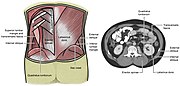 Lumbar triangle


