In anatomy, the atlas (C1) is the most superior (first) cervical vertebra of the spine and is located in the neck.

A spinal nerve is a mixed nerve, which carries motor, sensory, and autonomic signals between the spinal cord and the body. In the human body there are 31 pairs of spinal nerves, one on each side of the vertebral column. These are grouped into the corresponding cervical, thoracic, lumbar, sacral and coccygeal regions of the spine. There are eight pairs of cervical nerves, twelve pairs of thoracic nerves, five pairs of lumbar nerves, five pairs of sacral nerves, and one pair of coccygeal nerves. The spinal nerves are part of the peripheral nervous system.

The transverse abdominal muscle (TVA), also known as the transverse abdominis, transversalis muscle and transversus abdominis muscle, is a muscle layer of the anterior and lateral abdominal wall, deep to the internal oblique muscle. It is thought by most fitness instructors to be a significant component of the core.

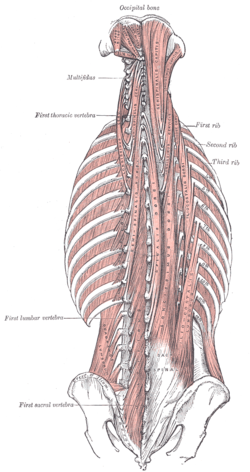
The multifidusmuscle consists of a number of fleshy and tendinous fasciculi, which fill up the groove on either side of the spinous processes of the vertebrae, from the sacrum to the axis. While very thin, the multifidus muscle plays an important role in stabilizing the joints within the spine. The multifidus is one of the transversospinales.

The rotatores muscles lie beneath the multifidus and are present in all spinal regions but are most prominent in the thoracic region; they are eleven in number on either side.

The semispinalis muscles are a group of three muscles belonging to the transversospinales. These are the semispinalis capitis, the semispinalis cervicis and the semispinalis thoracis.

The human back, also called the dorsum, is the large posterior area of the human body, rising from the top of the buttocks to the back of the neck. It is the surface of the body opposite from the chest and the abdomen. The vertebral column runs the length of the back and creates a central area of recession. The breadth of the back is created by the shoulders at the top and the pelvis at the bottom.

The occipital artery is a branch of the external carotid artery that provides arterial supply to the back of the scalp, sternocleidomastoid muscles, and deep muscles of the back and neck.

The erector spinae or spinal erectors is a set of muscles that straighten and rotate the back. The spinal erectors work together with the glutes to maintain stable posture standing or sitting.

Iliocostalis muscle is the muscle immediately lateral to the longissimus that is the nearest to the furrow that separates the epaxial muscles from the hypaxial. It lies very deep to the fleshy portion of the serratus posterior muscle. It laterally flexes the vertebral column to the same side.

The longissimus is the muscle lateral to the semispinalis muscles. It is the longest subdivision of the erector spinae muscles that extends forward into the transverse processes of the posterior cervical vertebrae.

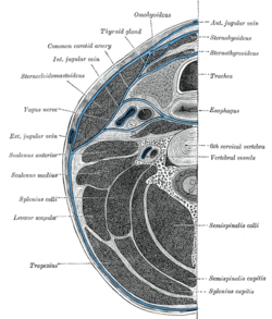
The deep cervical vein is the vena comitans of the deep cervical artery. The vein is formed in the suboccipital region by the convergence of communicating branches of the occipital vein, veins draining the suboccipital muscles, and veins from the venous plexuses that surround cervical nerves. The vein and corresponding artery then pass in between the semispinalis capitis muscle and the semispinalis colli muscle. The vein passes anterior-ward in between the transverse process of the 7th cervical vertebra and the neck of the first rib to terminate in the vertebral vein.

The crest of the ilium is the superior border of the wing of ilium and the superiolateral margin of the greater pelvis.
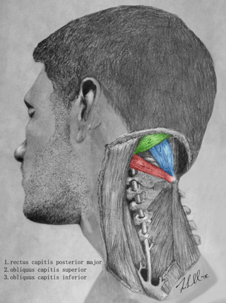
The suboccipital triangle is a region of the neck bounded by the following three muscles of the suboccipital group of muscles:

The deep cervical artery is an artery of the neck.
The posterior branches of thoracic nerves branch from the dorsal rami of the thoracic nerves.

The posterior branches of cervical nerves branch from the dorsal rami of the cervical nerves.
In kinesiology, core stability is a person's ability to stabilize their core. Stability, in this context, should be considered as an ability to control the position and movement of the core. Thus, if a person has greater core stability, they have a greater level of control over the position and movement of this area of their body. The body's core is frequently involved in aiding other movements of the body, such as running; thus it is known that improving core stability also improves a person's ability to perform these other movements.

In adult vertebrates, trunk muscles can be broadly divided into hypaxial muscles, which lie ventral to the horizontal septum of the vertebrae and epaxial muscles, which lie dorsal to the septum. Hypaxial muscles include some vertebral muscles, the diaphragm, the abdominal muscles, and all limb muscles. The serratus posterior inferior and serratus posterior superior are innervated by the ventral primary ramus and are hypaxial muscles. Epaxial muscles include other (dorsal) muscles associated with the vertebrae, ribs, and base of the skull. In humans, the erector spinae, the transversospinales, the splenius and suboccipital muscles are the only epaxial muscles.
This page is based on this
Wikipedia article Text is available under the
CC BY-SA 4.0 license; additional terms may apply.
Images, videos and audio are available under their respective licenses.