Subclavian nerve

Last updated
Subclavian nerve
Brachial plexus 2.svg
Diagram of the right brachial plexus. Subclavian nerve labelled at top right.
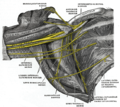
Gray808.png
The right brachial plexus with its short branches, viewed from in front. Subclavian nerve not visible.
Details
From Upper trunk (C5-C6) of brachial plexus
ToSometimes the accessory phrenic nerve
Innervates Subclavius muscle
Identifiers
Latin nervus subclavius
TA98 A14.2.03.013
TA2 6412
FMA 65280
Anatomical terms of neuroanatomy

The subclavian nerve, also known as the nerve to the subclavius, is a small branch of the upper trunk of the brachial plexus. It contains axons from C5 and C6. It innervates the subclavius muscle.
Contents

Anatomy

Origin

The subclavian nerve is a branch of the upper trunk of the brachial plexus. It contains axons derived from the ventral rami of the C5 and C6 cervical spinal nerves. [1]

The origin is situated within the posterior triangle of the neck. [2]

Course

Descending, it passes anterior to (the 3rd part of) the subclavian artery and vein. [2]

Variation

Accessory phrenic nerve

The subclavian nerve may issue a branch called the accessory phrenic nerve [3] [2] which innervates the diaphragm. [3] The accessory phrenic nerve may rather branch from the C4 or C6 segments or ansa cervicalis. [4] This nerve usually joins with the phrenic nerve before innervating the diaphragm, ventral to the subclavian vein. [5]

Function

The subclavian nerve innervates the subclavius muscle. [6]

Additional images

Related Research Articles

<span class="mw-page-title-main">Brachial plexus</span> Network of nerves

The brachial plexus is a network of nerves formed by the anterior rami of the lower four cervical nerves and first thoracic nerve. This plexus extends from the spinal cord, through the cervicoaxillary canal in the neck, over the first rib, and into the armpit, it supplies afferent and efferent nerve fibers to the chest, shoulder, arm, forearm, and hand.

<span class="mw-page-title-main">Spinal nerve</span> Nerve that carries signals between the spinal cord and the body

A spinal nerve is a mixed nerve, which carries motor, sensory, and autonomic signals between the spinal cord and the body. In the human body there are 31 pairs of spinal nerves, one on each side of the vertebral column. These are grouped into the corresponding cervical, thoracic, lumbar, sacral and coccygeal regions of the spine. There are eight pairs of cervical nerves, twelve pairs of thoracic nerves, five pairs of lumbar nerves, five pairs of sacral nerves, and one pair of coccygeal nerves. The spinal nerves are part of the peripheral nervous system.

<span class="mw-page-title-main">Axillary nerve</span> Nerve of the human body near the armpit

The axillary nerve or the circumflex nerve is a nerve of the human body, that originates from the brachial plexus at the level of the axilla (armpit) and carries nerve fibers from C5 and C6. The axillary nerve travels through the quadrangular space with the posterior circumflex humeral artery and vein to innervate the deltoid and teres minor.

<span class="mw-page-title-main">Phrenic nerve</span> Nerve controlling the diaphragm

The phrenic nerve is a mixed motor/sensory nerve that originates from the C3-C5 spinal nerves in the neck. The nerve is important for breathing because it provides exclusive motor control of the diaphragm, the primary muscle of respiration. In humans, the right and left phrenic nerves are primarily supplied by the C4 spinal nerve, but there is also a contribution from the C3 and C5 spinal nerves. From its origin in the neck, the nerve travels downward into the chest to pass between the heart and lungs towards the diaphragm.

<span class="mw-page-title-main">Long thoracic nerve</span> Large nerve

The long thoracic nerve is a branch of the brachial plexus derived from cervical nerves C5-C7 that innervates the serratus anterior muscle.

<span class="mw-page-title-main">Subclavian artery</span> Major arteries of the upper thorax, below the clavicle

In human anatomy, the subclavian arteries are paired major arteries of the upper thorax, below the clavicle. They receive blood from the aortic arch. The left subclavian artery supplies blood to the left arm and the right subclavian artery supplies blood to the right arm, with some branches supplying the head and thorax. On the left side of the body, the subclavian comes directly off the aortic arch, while on the right side it arises from the relatively short brachiocephalic artery when it bifurcates into the subclavian and the right common carotid artery.

<span class="mw-page-title-main">Infrahyoid muscles</span> Group of muscles

The infrahyoid muscles, or strap muscles, are a group of four pairs of muscles in the anterior (frontal) part of the neck. The four infrahyoid muscles are the sternohyoid, sternothyroid, thyrohyoid and omohyoid muscles.

<span class="mw-page-title-main">Cervical plexus</span> Network of nerves in the neck

The cervical plexus is a nerve plexus of the anterior rami of the first four cervical spinal nerves C1-C4. The cervical plexus provides motor innervation to some muscles of the neck, and the diaphragm; it provides sensory innervation to parts of the head, neck, and chest.

<span class="mw-page-title-main">Suprascapular nerve</span> Mixed nerve of the upper limb

The suprascapular nerve is a mixed nerve that branches from the upper trunk of the brachial plexus. It is derived from the ventral rami of cervical nerves C5-C6. It provides motor innervation to the supraspinatus muscle, and the infraspinatus muscle.

<span class="mw-page-title-main">Scalene muscles</span> Muscles on the sides of the neck

The scalene muscles are a group of three muscles on each side of the neck, identified as the anterior, the middle, and the posterior. They are innervated by the third to the eighth cervical spinal nerves (C3-C8).

<span class="mw-page-title-main">Nerve plexus</span> Network of nerve fibres

A nerve plexus is a plexus of intersecting nerves. A nerve plexus is composed of afferent and efferent fibers that arise from the merging of the anterior rami of spinal nerves and blood vessels. There are five spinal nerve plexuses, except in the thoracic region, as well as other forms of autonomic plexuses, many of which are a part of the enteric nervous system. The nerves that arise from the plexuses have both sensory and motor functions. These functions include muscle contraction, the maintenance of body coordination and control, and the reaction to sensations such as heat, cold, pain, and pressure. There are several plexuses in the body, including:

<span class="mw-page-title-main">Medial pectoral nerve</span>

The medial pectoral nerve is (typically) a branch of the medial cord of the brachial plexus and is derived from spinal nerve roots C8-T1. It provides motor innervation to the pectoralis minor muscle, and the lower half of the pectoralis major muscle. It runs along the inferior border of the pectoralis minor muscle.

<span class="mw-page-title-main">Thoracodorsal nerve</span> Nerve that supplies the latissimus dorsi

The thoracodorsal nerve is a nerve present in humans and other animals, also known as the middle subscapular nerve or the long subscapular nerve. It supplies the latissimus dorsi muscle.

<span class="mw-page-title-main">Posterior triangle of the neck</span> Region of the neck

The posterior triangle is a region of the neck.

<span class="mw-page-title-main">Lower subscapular nerve</span>

The lower subscapular nerve, also known as the inferior subscapular nerve, is the third branch of the posterior cord of the brachial plexus. It innervates the inferior portion of the subscapularis muscle and the teres major muscle.

<span class="mw-page-title-main">Upper subscapular nerve</span> Nerve of upper limb

The upper (superior) subscapular nerve is the first branch of the posterior cord of the brachial plexus. The upper subscapular nerve contains axons from the ventral rami of the C5 and C6 cervical spinal nerves. It innervates the superior portion of the subscapularis muscle. The inferior portion of the subscapularis is innervated by the lower subscapular nerve.

<span class="mw-page-title-main">Lateral pectoral nerve</span>

The lateral pectoral nerve arises from the lateral cord of the brachial plexus, and through it from the C5-7.

<span class="mw-page-title-main">Nerve point of neck</span> Aspect of human anatomy

The nerve point of the neck, also known as Erb's point, is a site at the upper trunk of the brachial plexus located 2–3 cm above the clavicle. It is named for Wilhelm Heinrich Erb. Taken together, there are six types of nerves that meet at this point.

<span class="mw-page-title-main">Ventral ramus of spinal nerve</span> Anterior division of a spinal nerve

The ventral ramus is the anterior division of a spinal nerve. The ventral rami supply the antero-lateral parts of the trunk and the limbs. They are mainly larger than the dorsal rami.

The upper (superior) trunk is part of the brachial plexus. It is formed by joining of the ventral rami of the fifth (C5) and sixth (C6) cervical nerves. The upper trunk divides into an anterior and posterior division.

References

  1. Rubin, Michael (28 September 2016). Netter's concise neuroanatomy. Netter, Frank H. (Frank Henry), 1906-1991 (Updated ed.). Philadelphia, PA. p. 310. ISBN   978-0-323-48091-8. OCLC   946698976.{{cite book}}: CS1 maint: location missing publisher (link)
  2. 1 2 3 Kadasne, D. K. (2009). Kadasne's Textbook of Anatomy (1st ed.). New Delhi: Jaypee Brothers Medical Publishers. ISBN   978-81-8448-455-7. OCLC   682534511.
  3. 1 2 Loukas, Marios; Kinsella, Christopher R.; Louis, Robert G.; Gandhi, Sagar; Curry, Brian (November 2006). "Surgical anatomy of the accessory phrenic nerve". The Annals of Thoracic Surgery. 82 (5): 1870–1875. doi:10.1016/j.athoracsur.2006.05.098. ISSN   1552-6259. PMID   17062263.
  4. Kayalioglu, Gulgun (2009-01-01), "Chapter 4 - The Spinal Nerves", in Watson, Charles; Paxinos, George; Kayalioglu, Gulgun (eds.), The Spinal Cord, San Diego: Academic Press, pp. 37–56, ISBN   978-0-12-374247-6 , retrieved 2022-06-14
  5. Sharma, Manish S.; Loukas, Marios; Spinner, Robert J. (2011). "Accessory phrenic nerve: A rarely discussed common variation with clinical implications". Clinical Anatomy. 24 (5): 671–673. doi:10.1002/ca.21142. PMID   21374726. S2CID   20001098.
  6. Drake, Richard L. (2005). Gray's anatomy for students. Wayne Vogl, Adam W. M. Mitchell, Henry Gray. Philadelphia: Elsevier / Churchill Livingstone. p. 645. ISBN   0-443-06612-4. OCLC   55139039.{{cite book}}: CS1 maint: date and year (link)