
The median nerve is a nerve in humans and other animals in the upper limb. It is one of the five main nerves originating from the brachial plexus.

The ulnar nerve is a nerve that runs near the ulna, one of the two long bones in the forearm. The ulnar collateral ligament of elbow joint is in relation with the ulnar nerve. The nerve is the largest in the human body unprotected by muscle or bone, so injury is common. This nerve is directly connected to the little finger, and the adjacent half of the ring finger, innervating the palmar aspect of these fingers, including both front and back of the tips, perhaps as far back as the fingernail beds.

The ulnar artery is the main blood vessel, with oxygenated blood, of the medial aspects of the forearm. It arises from the brachial artery and terminates in the superficial palmar arch, which joins with the superficial branch of the radial artery. It is palpable on the anterior and medial aspect of the wrist.

The tibial nerve is a branch of the sciatic nerve. The tibial nerve passes through the popliteal fossa to pass below the arch of soleus.

The flexor pollicis brevis is a muscle in the hand that flexes the thumb. It is one of three thenar muscles. It has both a superficial part and a deep part.

The flexor retinaculum is a fibrous band on the palmar side of the hand near the wrist. It arches over the carpal bones of the hands, covering them and forming the carpal tunnel.

The lateral cutaneous nerve of forearm is a sensory nerve representing the continuation of the musculocutaneous nerve beyond the lateral edge of the tendon of the biceps brachii muscle. The lateral cutaneous nerve provides sensory innervation to the skin of the lateral forearm. It pierces the deep fascia of forearm to enter the subcutaneous compartment before splitting into a volar branch and a dorsal branch.
The medial cutaneous nerve of the forearm is a sensory branch of the medial cord of the brachial plexus derived from the ventral rami of spinal nerves C8-T1. It provides sensory innervation to the skin of the medial forearm and skin overlying the olecranon. It descends through the (upper) arm within the brachial fascia alongside the basilic vein, then divides into an anterior branch and a posterior branch upon emerging from the brachial fascia; the two terminal branches travel as far distally as the wrist.
The medial brachial cutaneous nerve is a sensory branch of the medial cord of the brachial plexus derived from spinal nerves C8-T1. It provides sensory innervation to the medial arm. It descends accompanied by the basilic vein.
The intercostobrachial nerve is the name applied to the lateral cutaneous branch of the second intercostal nerve. It arises anterior to the long thoracic nerve. It provides sensory innervation to the skin of the axilla, and a variable region of the medial side of the upper arm.
The superficial branch of the radial nerve passes along the front of the radial side of the forearm to the commencement of its lower third. It is a sensory nerve.

The medial plantar nerve is the larger of the two terminal divisions of the tibial nerve, which accompanies the medial plantar artery.

The posterior cutaneous nerve of arm is a branch of the radial nerve that provides sensory innervation for much of the skin on the back of the arm. It arises in the axilla.

In the palm of the hand the median nerve is covered by the skin and the palmar aponeurosis, and rests on the tendons of the flexor muscles. Immediately after emerging from under the transverse carpal ligament the median nerve becomes enlarged and flattened and splits into a smaller, lateral, and a larger, medial portion.
In the palm of the hand, the median nerve is covered by the skin and the palmar aponeurosis, and rests on the tendons of the Flexor muscles. Immediately after emerging from under the transverse carpal ligament the median nerve becomes enlarged and flattened and splits into a smaller, lateral, and a larger, medial portion.

The lateral sural cutaneous nerve of the lumbosacral plexus supplies the skin on the posterior and lateral surfaces of the leg. The lateral sural cutaneous nerve originates from the common fibular nerve(L4-S2) and is the terminal branch of the common fibular nerve.
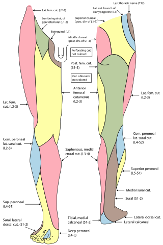
The anterior cutaneous branches of the femoral nerve consist of the following nerves: intermediate cutaneous nerve and medial cutaneous nerve.
Cutaneous innervation of the upper limbs is the nerve supply to areas of the skin of the upper limbs which are supplied by specific cutaneous nerves.

The posterior branches of cervical nerves branch from the dorsal rami of the cervical nerves.
Injuries to the arm, forearm or wrist area can lead to various nerve disorders. One such disorder is median nerve palsy. The median nerve controls the majority of the muscles in the forearm. It controls abduction of the thumb, flexion of hand at wrist, flexion of digital phalanx of the fingers, is the sensory nerve for the first three fingers, etc. Because of this major role of the median nerve, it is also called the eye of the hand. If the median nerve is damaged, the ability to abduct and oppose the thumb may be lost due to paralysis of the thenar muscles. Various other symptoms can occur which may be repaired through surgery and tendon transfers. Tendon transfers have been very successful in restoring motor function and improving functional outcomes in patients with median nerve palsy.


