|
| Part of a series of lists about |
| Human anatomy |
|---|
The following outline is provided as an overview of and topical guide to human anatomy:
Contents
- Essence of human anatomy
- Branches of human anatomy
- Anatomy of the human body
- General anatomy
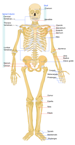
- Bones
- Joints
- Muscles
- Alimentary system
- Respiratory system
- Thoracic cavity
- Urinary system
- Genital systems
- Abdominopelvic cavity
- Endocrine glands
- Cardiovascular system
- Lymphoid system
- Nervous system
- Sense organs
- The integument
- History of human anatomy
- Organizations
- Anatomists
- See also
- External links
Human anatomy is the scientific study of the anatomy of the adult human. It is subdivided into gross anatomy and microscopic anatomy. Gross anatomy (also called topographical anatomy, regional anatomy, or anthropotomy) is the study of anatomical structures that can be seen by unaided vision. Microscopic anatomy is the study of minute anatomical structures assisted with microscopes, and includes histology (the study of the organization of tissues), and cytology (the study of cells).