Median cubital vein

Last updated
Median cubital vein
Gray574.png
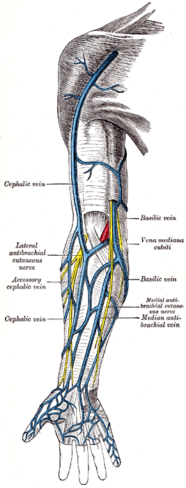
Superficial veins of the arm. The median cubital vein is labelled (in Latin) - Vena mediana cubiti.
Details
Source Cephalic vein
Drains to Basilic vein
Identifiers
Latin v. mediana cubiti,
v. intermedia cubiti
TA98 A12.3.08.019
TA2 4980
FMA 22963
Anatomical terminology

In human anatomy, the median cubital vein (or median basilic vein) is a superficial vein of the arm on the anterior aspect of the elbow. It classically connects the cephalic vein and the basilic vein. It is typically the most prominent superficial vein in the human body, and is visible when all other veins are hidden by fat or collapsed during a shock.

Contents

It is routinely used for venipuncture (taking blood) and as a site for an intravenous cannula. This is due to its particularly wide lumen, and its tendency to remain stationary upon needle insertion.

Structure

The median cubital vein is a superficial vein of the arm. [1] It lies on the anterior aspect of the elbow, [2] in the cubital fossa superficial to the bicipital aponeurosis. It bridges the cephalic vein and the basilic vein. [3]

The median cubital vein receives a number of tributaries from the anterior forearm. The median antebrachial vein may or may not drain into the median cubital vein. [3]

The median cubital vein issues a branch - the deep median vein - which pierces the fascial roof of the cubital fossa to join the brachial veins. [3]

Variations

The arrangement of the basilic, cephalic, median cubital, and median antebrachial veins exhibits a wide range of variations. Classically, the median cubital vein bridges the cephalic and basilic veins across the midline to form a H-pattern. However, in many cases, the median cubial vein is absent, instead replaced by a dominant median antebrachial vein that splits into intermediate cephalic and basilic veins to drain in the cephalic and basilic veins, respectively, forming a M-pattern. A number of other patterns exist, including ones where no anastomosis occurs between the basilic and cephalic veins, and ones where the median cubital vein is doubled. [4] When the median cubital vein is large, it transfers most or all blood from the cephalic vein to the basilic vein, so that the cephalic vein is either significantly diminished or altogether absent. [3]

Clinical significance

The median cubital vein becomes prominent when pressure is applied to upstream veins as venous blood builds up. [5]

The median cubital vein is routinely used for phlebotomy and venipuncture (taking blood), and as a site for an intravenous cannula. [6] This is due to its particularly wide lumen, and its tendency to remain stationary upon needle insertion. [6] It becomes prominent when pressure is applied upstream, which makes needle insertion easier. [5] Such pressure is created using a tourniquet. [5]

Additional images

See also

Related Research Articles

<span class="mw-page-title-main">Arm</span> Proximal part of the free upper limb between the shoulder and the elbow

In human anatomy, the arm refers to the upper limb in common usage, although academically the term specifically means the upper arm between the glenohumeral joint and the elbow joint. The distal part of the upper limb between the elbow and the radiocarpal joint is known as the forearm or "lower" arm, and the extremity beyond the wrist is the hand.

<span class="mw-page-title-main">Venipuncture</span> Process of obtaining intravenous access

In medicine, venipuncture or venepuncture is the process of obtaining intravenous access for the purpose of venous blood sampling or intravenous therapy. In healthcare, this procedure is performed by medical laboratory scientists, medical practitioners, some EMTs, paramedics, phlebotomists, dialysis technicians, and other nursing staff. In veterinary medicine, the procedure is performed by veterinarians and veterinary technicians.

The forearm is the region of the upper limb between the elbow and the wrist. The term forearm is used in anatomy to distinguish it from the arm, a word which is used to describe the entire appendage of the upper limb, but which in anatomy, technically, means only the region of the upper arm, whereas the lower "arm" is called the forearm. It is homologous to the region of the leg that lies between the knee and the ankle joints, the crus.

<span class="mw-page-title-main">Great saphenous vein</span> Large, subcutaneous, superficial vein of the leg

The great saphenous vein (GSV) or long saphenous vein is a large, subcutaneous, superficial vein of the leg. It is the longest vein in the body, running along the length of the lower limb, returning blood from the foot, leg and thigh to the deep femoral vein at the femoral triangle.

<span class="mw-page-title-main">Ulnar nerve</span> Nerve which runs near the ulna bone

The ulnar nerve is a nerve that runs near the ulna, one of the two long bones in the forearm. The ulnar collateral ligament of elbow joint is in relation with the ulnar nerve. The nerve is the largest in the human body unprotected by muscle or bone, so injury is common. This nerve is directly connected to the little finger, and the adjacent half of the ring finger, innervating the palmar aspect of these fingers, including both front and back of the tips, perhaps as far back as the fingernail beds.

<span class="mw-page-title-main">Popliteal vein</span> Large leg vein

The popliteal vein is a vein of the lower limb. It is formed from the anterior tibial vein and the posterior tibial vein. It travels medial to the popliteal artery, and becomes the femoral vein. It drains blood from the leg. It can be assessed using medical ultrasound. It can be affected by popliteal vein entrapment.

<span class="mw-page-title-main">Cephalic vein</span> Large blood vessel in the arm

In human anatomy, the cephalic vein is a superficial vein in the arm. It originates from the radial end of the dorsal venous network of hand, and ascends along the radial (lateral) side of the arm before emptying into the axillary vein. At the elbow, it communicates with the basilic vein via the median cubital vein.

<span class="mw-page-title-main">Cubital fossa</span> The human elbow pit

The cubital fossa, antecubital fossa, chelidon, or inside of elbow is the area on the anterior side of the upper part between the arm and forearm of a human or other hominid animals. It lies anteriorly to the elbow (antecubital) when in standard anatomical position. The cubital fossa is a triangular area having three borders.

<span class="mw-page-title-main">Superficial vein</span> Veins close to the surface of the body

Superficial veins are veins that are close to the surface of the body, as opposed to deep veins, which are far from the surface.

<span class="mw-page-title-main">Basilic vein</span> Large blood vessel in the arm

The basilic vein is a large superficial vein of the upper limb that helps drain parts of the hand and forearm. It originates on the medial (ulnar) side of the dorsal venous network of the hand and travels up the base of the forearm, where its course is generally visible through the skin as it travels in the subcutaneous fat and fascia lying superficial to the muscles. The basilic vein terminates by uniting with the brachial veins to form the axillary vein.

<span class="mw-page-title-main">Ulnar veins</span> Large blood vessels of the forearm

The ulnar veins are venae comitantes of the ulnar artery. They drain the superficial venous palmar arch. They arise in the hand and terminate by uniting with the radial veins to form the brachial veins. They mostly drain the medial aspect of the forearm. They receive the venae comitantes of the anterior and posterior interosseous arteries near the elbow, as well as a large branch from the median cubital vein. The ulnar veins are larger than the radial veins.

The semimembranosus muscle is the most medial of the three hamstring muscles in the thigh. It is so named because it has a flat tendon of origin. It lies posteromedially in the thigh, deep to the semitendinosus muscle. It extends the hip joint and flexes the knee joint.

<span class="mw-page-title-main">Superior epigastric vein</span> Blood vessel

In human anatomy, the superior epigastric veins are two or more venae comitantes which accompany either superior epigastric artery before emptying into the internal thoracic vein. They participate in the drainage of the superior surface of the diaphragm.

<span class="mw-page-title-main">Inferior ophthalmic vein</span> Vein of the orbit around the eye

The inferior ophthalmic vein is a vein of the orbit that - together with the superior ophthalmic vein - represents the principal drainage system of the orbit. It begins from a venous network in the front of the orbit, then passes backwards through the lower orbit. It drains several structures of the orbit. It may end by splitting into two branches, one draining into the pterygoid venous plexus and the other ultimately into the cavernous sinus.

<span class="mw-page-title-main">Incisive foramen</span> Opening of the incisive canals on the hard palate immediately behind the incisor teeth

In the human mouth, the incisive foramen is the opening of the incisive canals on the hard palate immediately behind the incisor teeth. It gives passage to blood vessels and nerves. The incisive foramen is situated within the incisive fossa of the maxilla.

<span class="mw-page-title-main">Retromandibular vein</span> Major face vein

The retromandibular vein is a major vein of the face. It is formed within the parotid gland by the confluence of the maxillary vein, and superficial temporal vein. It descends in the gland and splits into two branches upon emerging from the gland. Its anterior branch then joins the (anterior) facial vein forming the common facial vein, while its posterior branch joins the posterior auricular vein forming the external jugular vein.

<span class="mw-page-title-main">Bicipital aponeurosis</span> Distal end of the biceps muscle

The bicipital aponeurosis is a broad aponeurosis of the biceps brachii, which is located in the cubital fossa of the elbow. It separates superficial from deep structures in much of the fossa.

<span class="mw-page-title-main">Median antebrachial vein</span> Vein draining the venous plexus on the palmar surface of the hand

The median antebrachial vein is a superficial vein of the (anterior) forearm. It arises from - and drains - the superficial palmar venous arch, ascending superficially along the anterior forearm before terminating by draining into either the basilic vein and/or median cubital vein; it may bifurcate distal to the elbow and proceed to drain into both aforementioned veins. A bifurcation of the median antebrachial vein produces the (medial) intermediate basilic vein and the (lateral) intermediate cephalic vein; the two veins produced by such a split may replace the median cubital vein.

<span class="mw-page-title-main">Sphenoparietal sinus</span>

The sphenoparietal sinus is a paired dural venous sinus situated along the posterior edge of the lesser wing of either sphenoid bone. It drains into the cavernous sinus.

<span class="mw-page-title-main">Superficial venous palmar arch</span>

The superficial palmar venous arch consists of a pair of venae comitantes accompanying the superficial palmar arch. It receives the common palmar digital veins. It drains into the superficial ulnar radial and superficial radial veins, and the median antebrachial vein.

References

  1. Standring, Susan (2016). Gray's anatomy: the anatomical basis of clinical practice (41 ed.). Elsevier Limited. pp. 837–861. ISBN   978-0-7020-5230-9.
  2. Standring, Susan (2020). Gray's Anatomy: The Anatomical Basis of Clinical Practice (42th ed.). New York. p. 612. ISBN   978-0-7020-7707-4. OCLC   1201341621.{{cite book}}: CS1 maint: location missing publisher (link)
  3. 1 2 3 4 Hunter, James P.; Barlow, Adam D.; Nicholson, Michael L. (2014). "5 - Access for Renal Replacement Therapy". Kidney Transplantation–Principles and Practice (7th ed.). Saunders. pp. 72–90. doi:10.1016/B978-1-4557-4096-3.00005-2. ISBN   978-1-4557-4096-3.
  4. 1 2 Pires, L.; Ráfare, A. L.; Peixoto, B. U.; Pereira, T. O. J. S.; Pinheiro, D. M. M.; Siqueira, M. E. B.; Vaqueiro, R. D.; de Paula, R. C.; Babinski, M. A.; Chagas, C. A. A. (2018-06-01). "The venous patterns of the cubital fossa in subjects from Brazil". Morphologie. 102 (337): 78–82. doi:10.1016/j.morpho.2018.02.001. ISSN   1286-0115. PMID   29625795. S2CID   4662703.
  5. 1 2 3 Sadrzadeh, Hossein; Baskin, Leland; Kline, Gregory (2017). "1 - Variables affecting endocrine tests results, errors prevention and mitigation". Endocrine Biomarkers - Clinicians and Clinical Chemists in Partnership. Elsevier. pp. 1–40. doi:10.1016/B978-0-12-803412-5.00001-X. ISBN   978-0-12-803412-5.
  6. 1 2 Lew, K. (2012-01-01), Pawliszyn, Janusz (ed.), "3.05 - Blood Sample Collection and Handling", Comprehensive Sampling and Sample Preparation, Oxford: Academic Press, pp. 95–121, doi:10.1016/b978-0-12-381373-2.00068-5, ISBN   978-0-12-381374-9 , retrieved 2020-11-17