Accessory cephalic vein

Last updated
Accessory cephalic vein
Gray574.png
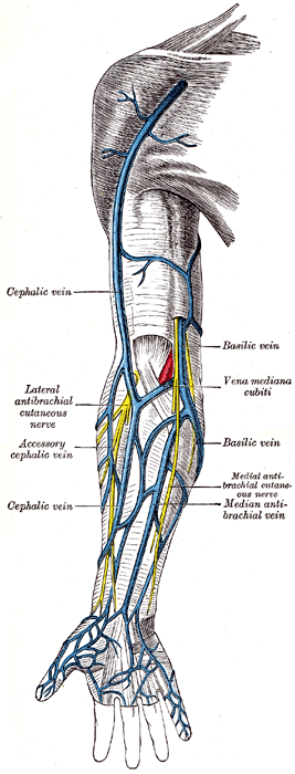
Superficial veins of the upper limb (accessory cephalic vein labeled at center left)
Details
Source Dorsal venous network of hand
Drains to Cephalic vein
Identifiers
Latin vena cephalica accessoria
TA98 A12.3.08.017
TA2 4966
FMA 22970
Anatomical terminology

The accessory cephalic vein is a variable vein that passes along the radial border of the forearm to join the cephalic vein [1] distal/inferior to the elbow. It may arise from a dorsal forearm venous plexus, or from the ulnar/medial side of the dorsal venous network of hand. [2] In some cases the accessory cephalic springs from the cephalic above the wrist and joins it again higher up. A large oblique branch frequently connects the basilic and cephalic veins on the back of the forearm.[ citation needed ]

See also

Related Research Articles

<span class="mw-page-title-main">Arm</span> Proximal part of the free upper limb between the shoulder and the elbow

In human anatomy, the arm refers to the upper limb in common usage, although academically the term specifically means the upper arm between the glenohumeral joint and the elbow joint. The distal part of the upper limb between the elbow and the radiocarpal joint is known as the forearm or "lower" arm, and the extremity beyond the wrist is the hand.

<span class="mw-page-title-main">Anatomical snuffbox</span> Indent on back of hand between tendons

The anatomical snuff box or snuffbox or foveola radialis is a triangular deepening on the radial, dorsal aspect of the hand—at the level of the carpal bones, specifically, the scaphoid and trapezium bones forming the floor. The name originates from the use of this surface for placing and then sniffing powdered tobacco, or "snuff." It is sometimes referred to by its French name tabatière.

The forearm is the region of the upper limb between the elbow and the wrist. The term forearm is used in anatomy to distinguish it from the arm, a word which is used to describe the entire appendage of the upper limb, but which in anatomy, technically, means only the region of the upper arm, whereas the lower "arm" is called the forearm. It is homologous to the region of the leg that lies between the knee and the ankle joints, the crus.

<span class="mw-page-title-main">Great saphenous vein</span> Large, subcutaneous, superficial vein of the leg

The great saphenous vein (GSV) or long saphenous vein is a large, subcutaneous, superficial vein of the leg. It is the longest vein in the body, running along the length of the lower limb, returning blood from the foot, leg and thigh to the deep femoral vein at the femoral triangle.

<span class="mw-page-title-main">Azygos vein</span> Human blood vessel by the spine

The azygos vein is a vein running up the right side of the thoracic vertebral column draining itself towards the superior vena cava. It connects the systems of superior vena cava and inferior vena cava and can provide an alternative path for blood to the right atrium when either of the venae cavae is blocked.

<span class="mw-page-title-main">Radial artery</span> Large forearm artery

In human anatomy, the radial artery is the main artery of the lateral aspect of the forearm.

<span class="mw-page-title-main">External jugular vein</span> Large blood vessel

The external jugular vein receives the greater part of the blood from the exterior of the cranium and the deep parts of the face, being formed by the junction of the posterior division of the retromandibular vein with the posterior auricular vein.

<span class="mw-page-title-main">Cephalic vein</span> Large blood vessel in the arm

In human anatomy, the cephalic vein is a superficial vein in the arm. It originates from the radial end of the dorsal venous network of hand, and ascends along the radial (lateral) side of the arm before emptying into the axillary vein. At the elbow, it communicates with the basilic vein via the median cubital vein.

<span class="mw-page-title-main">Cubital fossa</span> The human elbow pit

The cubital fossa, super pit, chelidon or inside of elbow is the area on the anterior side of the upper part between the arm and forearm of a human or other hormid animals. It lies anteriorly to the elbow when in standard anatomical position.

In human anatomy, the median cubital vein is a superficial vein of the arm on the anterior aspect of the elbow. It classically connects the cephalic vein and the basilic vein.

<span class="mw-page-title-main">Basilic vein</span> Large blood vessel in the arm

The basilic vein is a large superficial vein of the upper limb that helps drain parts of the hand and forearm. It originates on the medial (ulnar) side of the dorsal venous network of the hand and travels up the base of the forearm, where its course is generally visible through the skin as it travels in the subcutaneous fat and fascia lying superficial to the muscles. The basilic vein terminates by uniting with the brachial veins to form the axillary vein.

<span class="mw-page-title-main">Lateral cutaneous nerve of forearm</span>

The lateral cutaneous nerve of forearm is a sensory nerve representing the continuation of the musculocutaneous nerve beyond the lateral edge of the tendon of the biceps brachii muscle. The lateral cutaneous nerve provides sensory innervation to the skin of the lateral forearm. It pierces the deep fascia of forearm to enter the subcutaneous compartment before splitting into a volar branch and a dorsal branch.

<span class="mw-page-title-main">Medial cutaneous nerve of forearm</span>

The medial cutaneous nerve of the forearm is a sensory branch of the medial cord of the brachial plexus derived from the ventral rami of spinal nerves C8-T1. It provides sensory innervation to the skin of the medial forearm and skin overlying the olecranon. It descends through the (upper) arm within the brachial fascia alongside the basilic vein, then divides into an anterior branch and a posterior branch upon emerging from the brachial fascia; the two terminal branches travel as far distally as the wrist.

<span class="mw-page-title-main">Posterior cutaneous nerve of forearm</span>

The posterior cutaneous nerve of forearm is a nerve found in humans and other animals. It is also known as the dorsal antebrachial cutaneous nerve, the external cutaneous branch of the musculospiral nerve, and the posterior antebrachial cutaneous nerve. It is a cutaneous nerve of the forearm.

<span class="mw-page-title-main">Median antebrachial vein</span> Vein draining the venous plexus on the palmar surface of the hand

The median antebrachial vein is a superficial vein of the (anterior) forearm. It arises from - and drains - the superficial palmar venous arch, ascending superficially along the anterior forearm before terminating by draining into either the basilic vein and/or median cubital vein; it may bifurcate distal to the elbow and proceed to drain into both aforementioned veins. A bifurcation of the median antebrachial vein produces the (medial) intermediate basilic vein and the (lateral) intermediate cephalic vein; the two veins produced by such a split may replace the median cubital vein.

<span class="mw-page-title-main">Cerebellar veins</span> Veins that drain the cerebellum

The cerebellar veins are veins which drain the cerebellum. They consist of the superior cerebellar veins and the inferior cerebellar veins. The superior cerebellar veins drain to the straight sinus and the internal cerebral veins. The inferior cerebellar veins drain to the transverse sinus, the superior petrosal sinus, and the occipital sinus.

<span class="mw-page-title-main">Dorsal venous network of hand</span> Veins on the back of the hand

The dorsal venous network of the hand is a venous network on the dorsum (backside) of hand. It is formed by the dorsal metacarpal veins, a dorsal digital vein from the radial side of the index finger and one from the ulnar side of the little finger, and both dorsal digital veins of the thumb. The venous network gives rise to the cephalic vein and the basilic vein; an accessory cephalic vein may arise from it as well.

On the dorsum of the foot the dorsal digital veins receive, in the clefts between the toes, the intercapitular veins from the plantar venous arch and join to form short common digital veins which unite across the distal ends of the metatarsal bones in a dorsal venous arch.

<span class="mw-page-title-main">Vesical venous plexus</span>

The vesical venous plexus is a venous plexus situated at the fundus of the urinary bladder. It collects venous blood from the urinary bladder in both sexes, from the accessory sex glands in males, and from the corpora cavernosa of clitoris in females. It drains into the internal iliac veins via several vesical veins.

On the dorsum of the foot the dorsal digital veins receive, in the clefts between the toes, the intercapitular veins from the plantar venous arch and join to form short common digital veins.

References

PD-icon.svgThis article incorporates text in the public domain from page 662 of the 20th edition of Gray's Anatomy (1918)

  1. http://dictionary.reference.com/browse/accessory+cephalic+vein (10/15/10)
  2. Standring, Susan (2020). Gray's Anatomy: The Anatomical Basis of Clinical Practice (42th ed.). New York. p. 1464.e104. ISBN   978-0-7020-7707-4. OCLC   1201341621.{{cite book}}: CS1 maint: location missing publisher (link)