Veins are blood vessels in the circulatory system of humans and most other animals that carry blood towards the heart. Most veins carry deoxygenated blood from the tissues back to the heart; exceptions are those of the pulmonary and fetal circulations which carry oxygenated blood to the heart. In the systemic circulation, arteries carry oxygenated blood away from the heart, and veins return deoxygenated blood to the heart, in the deep veins.

The circulatory system is a system of organs that includes the heart, blood vessels, and blood which is circulated throughout the entire body of a human or other vertebrate. It includes the cardiovascular system, or vascular system, that consists of the heart and blood vessels. The circulatory system has two divisions, a systemic circulation or circuit, and a pulmonary circulation or circuit. Some sources use the terms cardiovascular system and vascular system interchangeably with circulatory system.

The great saphenous vein (GSV) or long saphenous vein is a large, subcutaneous, superficial vein of the leg. It is the longest vein in the body, running along the length of the lower limb, returning blood from the foot, leg and thigh to the deep femoral vein at the femoral triangle.

The great cerebral vein is one of the large blood vessels in the skull draining the cerebrum of the brain. It is also known as the vein of Galen, named for its discoverer, the Greek physician Galen.
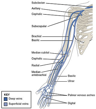
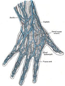
In human anatomy, the cephalic vein is a superficial vein in the arm. It originates from the radial end of the dorsal venous network of hand, and ascends along the radial (lateral) side of the arm before emptying into the axillary vein. At the elbow, it communicates with the basilic vein via the median cubital vein.
The small saphenous vein is a relatively large superficial vein of the posterior leg.
The dorsal venous arch of the foot is a superficial vein that connects the small saphenous vein and the great saphenous vein. Anatomically, it is defined by where the dorsal veins of the first and fifth digit, respectively, meet the great saphenous vein and small saphenous vein.

The anterior tibial vein is a vein in the lower leg.

The arachnoid mater is one of the three meninges, the protective membranes that cover the brain and spinal cord. It is so named because of its resemblance to a spider web. The arachnoid mater is a derivative of the neural crest mesoectoderm in the embryo.

The vitelline veins are veins that drain blood from the yolk sac and the gut tube during gestation.
The accessory cephalic vein is a variable vein that passes along the radial border of the forearm to join the cephalic vein distal/inferior to the elbow. It may arise from a dorsal forearm venous plexus, or from the ulnar/medial side of the dorsal venous network of hand. In some cases the accessory cephalic springs from the cephalic above the wrist and joins it again higher up. A large oblique branch frequently connects the basilic and cephalic veins on the back of the forearm.

The cerebellar veins are veins which drain the cerebellum. They consist of the superior cerebellar veins and the inferior cerebellar veins. The superior cerebellar veins drain to the straight sinus and the internal cerebral veins. The inferior cerebellar veins drain to the transverse sinus, the superior petrosal sinus, and the occipital sinus.

The dorsal venous network of the hand is a venous network on the dorsum (backside) of hand. It is formed by the dorsal metacarpal veins, a dorsal digital vein from the radial side of the index finger and one from the ulnar side of the little finger, and both dorsal digital veins of the thumb. The venous network gives rise to the cephalic vein and the basilic vein; an accessory cephalic vein may arise from it as well.
On the dorsum of the foot the dorsal digital veins receive, in the clefts between the toes, the intercapitular veins from the plantar venous arch and join to form short common digital veins which unite across the distal ends of the metatarsal bones in a dorsal venous arch.
The pudendal venous plexus lies behind the arcuate pubic ligament and the lower part of the pubic symphysis, and in front of the bladder and prostate. Its chief tributary is the deep dorsal vein of the penis, but it also receives branches from the front of the bladder and prostate. It communicates with the vesical venous plexus and with the internal pudendal vein and drains into the vesical and hypogastric veins.
The palmar digital veins on each finger are connected to the dorsal digital veins by oblique intercapitular veins. They drain into a venous plexus which is situated over the thenar and hypothenar eminences and across the front of the wrist.

Anterior spinal veins are veins that receive blood from the anterior spinal cord.
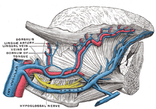
The dorsal lingual veins are some of the lingual veins. They provide venous drainage to the dorsum of the tongue, and the sides of the tongue. Between the hyoglossus and genioglossus, dorsal lingual veins unite with those lingual veins that are venae comitantes of the lingual artery; these consolidated lingual veins then empty into the internal jugular vein proximal to the greater cornu of hyoid bone.

Heart development, also known as cardiogenesis, refers to the prenatal development of the heart. This begins with the formation of two endocardial tubes which merge to form the tubular heart, also called the primitive heart tube. The heart is the first functional organ in vertebrate embryos.
This page is based on this
Wikipedia article Text is available under the
CC BY-SA 4.0 license; additional terms may apply.
Images, videos and audio are available under their respective licenses.