Basilic vein

Last updated

Basilic vein
2134 Thoracic Upper Limb Veins.jpg
Veins of the upper limb
Sobo 1909 597.png
The most frequent variations of the veins of the forearm (schematic).
Details
Source Dorsal venous network of hand
Drains to Axillary vein, median cubital vein
Identifiers
Latin vena basilica
TA98 A12.3.08.018
TA2 4979
FMA 22908
Anatomical terminology

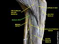
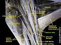
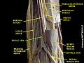
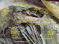
The basilic vein is a large superficial vein of the upper limb that helps drain parts of the hand and forearm. [1] It originates on the medial (ulnar) side of the dorsal venous network of the hand and travels up the base of the forearm, where its course is generally visible through the skin as it travels in the subcutaneous fat and fascia lying superficial to the muscles. The basilic vein terminates by uniting with the brachial veins to form the axillary vein.[ citation needed ]

Contents

Anatomy

Course

As it ascends the medial side of the biceps in the arm proper (between the elbow and shoulder), the basilic vein normally perforates the brachial fascia (deep fascia) superior to the medial epicondyle, or even as high as mid-arm.[ citation needed ]

Tributaries and anastomoses

Near the region anterior to the cubital fossa (in the bend of the elbow joint), the basilic vein usually communicates with the cephalic vein (the other large superficial vein of the upper extremity) via the median cubital vein. The layout of superficial veins in the forearm is highly variable from person to person, and there is a profuse network of unnamed superficial veins that the basilic vein communicates with.[ citation needed ]

Around the inferior border of the teres major muscle and just proximal to the basilic vein's termination, the anterior and posterior circumflex humeral veins drain into it.[ citation needed ]

Clinical significance

Venipuncture

Along with other superficial veins in the forearm, the basilic vein is an acceptable site for venipuncture. Nevertheless, IV nurses sometimes refer to the basilic vein as the "virgin vein", since with the arm typically supinated during phlebotomy the basilic vein below the elbow becomes awkward to access, and is therefore infrequently used.[ citation needed ]

Venous grafts

Vascular surgeons sometimes utilize the basilic vein to create an AV (arteriovenous) fistula or AV graft for hemodialysis access in patients with kidney failure. [2]

Additional images

See also

Related Research Articles

<span class="mw-page-title-main">Arm</span> Proximal part of the free upper limb between the shoulder and the elbow

In human anatomy, the arm refers to the upper limb in common usage, although academically the term specifically means the upper arm between the glenohumeral joint and the elbow joint. The distal part of the upper limb between the elbow and the radiocarpal joint is known as the forearm or "lower" arm, and the extremity beyond the wrist is the hand.

<span class="mw-page-title-main">Brachioradialis</span> Muscle of the upper limb

The brachioradialis is a muscle of the forearm that flexes the forearm at the elbow. It is also capable of both pronation and supination, depending on the position of the forearm. It is attached to the distal styloid process of the radius by way of the brachioradialis tendon, and to the lateral supracondylar ridge of the humerus.

<span class="mw-page-title-main">Radial nerve</span> Nerve in the human body that supplies the posterior portion of the upper limb

The radial nerve is a nerve in the human body that supplies the posterior portion of the upper limb. It innervates the medial and lateral heads of the triceps brachii muscle of the arm, as well as all 12 muscles in the posterior osteofascial compartment of the forearm and the associated joints and overlying skin.

<span class="mw-page-title-main">Brachial artery</span> Large blood vessel

The brachial artery is the major blood vessel of the (upper) arm. It is the continuation of the axillary artery beyond the lower margin of teres major muscle. It continues down the ventral surface of the arm until it reaches the cubital fossa at the elbow. It then divides into the radial and ulnar arteries which run down the forearm. In some individuals, the bifurcation occurs much earlier and the ulnar and radial arteries extend through the upper arm. The pulse of the brachial artery is palpable on the anterior aspect of the elbow, medial to the tendon of the biceps, and, with the use of a stethoscope and sphygmomanometer, often used to measure the blood pressure.

<span class="mw-page-title-main">Median nerve</span> Nerve of the upper limb

The median nerve is a nerve in humans and other animals in the upper limb. It is one of the five main nerves originating from the brachial plexus.

<span class="mw-page-title-main">Brachialis muscle</span> Flexor muscle in the upper arm

The brachialis is a muscle in the upper arm that flexes the elbow. It lies beneath the biceps brachii, and makes up part of the floor of the region known as the cubital fossa. It originates from the anterior aspect of the distal humerus; it inserts onto the tuberosity of the ulna. It is innervated by the musculocutaneous nerve, and commonly also receives additional innervation from the radial nerve. The brachialis is the prime mover of elbow flexion generating about 50% more power than the biceps.

The forearm is the region of the upper limb between the elbow and the wrist. The term forearm is used in anatomy to distinguish it from the arm, a word which is used to describe the entire appendage of the upper limb, but which in anatomy, technically, means only the region of the upper arm, whereas the lower "arm" is called the forearm. It is homologous to the region of the leg that lies between the knee and the ankle joints, the crus.

<span class="mw-page-title-main">Ulnar nerve</span> Nerve which runs near the ulna bone

In human anatomy, the ulnar nerve is a nerve that runs near the ulna bone. The ulnar collateral ligament of elbow joint is in relation with the ulnar nerve. The nerve is the largest in the human body unprotected by muscle or bone, so injury is common. This nerve is directly connected to the little finger, and the adjacent half of the ring finger, innervating the palmar aspect of these fingers, including both front and back of the tips, perhaps as far back as the fingernail beds.

<span class="mw-page-title-main">Upper limb</span> Consists of the arm, forearm, and hand

The upper limbs or upper extremities are the forelimbs of an upright-postured tetrapod vertebrate, extending from the scapulae and clavicles down to and including the digits, including all the musculatures and ligaments involved with the shoulder, elbow, wrist and knuckle joints. In humans, each upper limb is divided into the arm, forearm and hand, and is primarily used for climbing, lifting and manipulating objects.

<span class="mw-page-title-main">Cephalic vein</span> Large blood vessel in the arm

In human anatomy, the cephalic vein is a superficial vein in the arm. It originates from the radial end of the dorsal venous network of hand, and ascends along the radial (lateral) side of the arm before emptying into the axillary vein. At the elbow, it communicates with the basilic vein via the median cubital vein.

<span class="mw-page-title-main">Cubital fossa</span> The human elbow pit

The cubital fossa, chelidon or inside of elbow is the area on the anterior side of the upper part between the arm and forearm of a human or other hormid animals. It lies anteriorly to the elbow when in standard anatomical position.

In human anatomy, the median cubital vein is a superficial vein of the arm on the anterior aspect of the elbow. It classically connects the cephalic vein and the basilic vein.

<span class="mw-page-title-main">Ulnar artery</span> Artery of the forearm

The ulnar artery is the main blood vessel, with oxygenated blood, of the medial aspects of the forearm. It arises from the brachial artery and terminates in the superficial palmar arch, which joins with the superficial branch of the radial artery. It is palpable on the anterior and medial aspect of the wrist.

<span class="mw-page-title-main">Ulnar veins</span> Large blood vessels of the forearm

The ulnar veins are venae comitantes of the ulnar artery. They drain the superficial venous palmar arch. They arise in the hand and terminate by uniting with the radial veins to form the brachial veins. They mostly drain the medial aspect of the forearm. They receive the venae comitantes of the anterior and posterior interosseous arteries near the elbow, as well as a large branch from the median cubital vein. The ulnar veins are larger than the radial veins.

The pronator teres is a muscle that, along with the pronator quadratus, serves to pronate the forearm. It has two origins, at the medial humeral supracondylar ridge and the ulnar tuberosity, and inserts near the middle of the radius.

<span class="mw-page-title-main">Gracilis muscle</span> Most superficial muscle on the medial side of the thigh

The gracilis muscle is the most superficial muscle on the medial side of the thigh. It is thin and flattened, broad above, narrow and tapering below.

<span class="mw-page-title-main">Lateral cutaneous nerve of forearm</span>

The lateral cutaneous nerve of forearm is a sensory nerve representing the continuation of the musculocutaneous nerve beyond the lateral edge of the tendon of the biceps brachii muscle. The lateral cutaneous nerve provides sensory innervation to the skin of the lateral forearm. It pierces the deep fascia of forearm to enter the subcutaneous compartment before splitting into a volar branch and a dorsal branch.

<span class="mw-page-title-main">Medial cutaneous nerve of forearm</span>

The medial cutaneous nerve of the forearm is a sensory branch of the medial cord of the brachial plexus derived from the ventral rami of spinal nerves C8-T1. It provides sensory innervation to the skin of the medial forearm and skin overlying the olecranon. It descends through the (upper) arm within the brachial fascia alongside the basilic vein, then divides into an anterior branch and a posterior branch upon emerging from the brachial fascia; the two terminal branches travel as far distally as the wrist.

<span class="mw-page-title-main">Intercostobrachial nerve</span>

The intercostobrachial nerve is the name applied to the lateral cutaneous branch of the second intercostal nerve. It arises anterior to the long thoracic nerve. It provides sensory innervation to the skin of the axilla, and a variable region of the medial side of the upper arm.

<span class="mw-page-title-main">Elbow</span> Joint between the upper and lower parts of the arm

The elbow is the region between the upper arm and the forearm that surrounds the elbow joint. The elbow includes prominent landmarks such as the olecranon, the cubital fossa, and the lateral and the medial epicondyles of the humerus. The elbow joint is a hinge joint between the arm and the forearm; more specifically between the humerus in the upper arm and the radius and ulna in the forearm which allows the forearm and hand to be moved towards and away from the body. The term elbow is specifically used for humans and other primates, and in other vertebrates it is not used. In those cases, forelimb plus joint is used.

References

  1. Doyle, James R. (2003). Surgical anatomy of the hand and upper extremity. Michael J. Botte. Philadelphia: Lippincott Williams & Wilkins. ISBN   978-0-397-51725-1. OCLC   421140820.
  2. Kakkos, S. K.; Haddad, G. K.; Weaver, M. R.; Haddad, R. K.; Scully, M. M. (May 1, 2010). "Basilic Vein Transposition: What is the Optimal Technique?". European Journal of Vascular and Endovascular Surgery. 39 (5): 612–619. doi: 10.1016/j.ejvs.2010.01.006 . ISSN   1078-5884.