Great auricular nerve

Last updated
Great auricular nerve
Gray805.png
The nerves of the scalp, face, and side of neck. (Great auricular visible below ear.)
Gray804.png
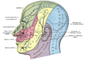
Plan of the cervical plexus. (Great auricular labeled at top center.)
Details
From cervical plexus (C2-C3)
Innervatessensation of inferior part of auricle and parotid region of the face.
Identifiers
Latin nervus auricularis magnus
TA98 A14.2.02.018
TA2 6385
FMA 6872
Anatomical terms of neuroanatomy

The great auricular nerve is a cutaneous (sensory) nerve of the head. It originates from the second and third cervical (spinal) nerves (C2-C3) of the cervical plexus. It provides sensory innervation to the skin over the parotid gland and the mastoid process, parts of the outer ear, and to the parotid gland and its fascia.

Contents

Pain resulting from parotitis is caused by an impingement on the great auricular nerve.

Structure

The great auricular nerve is the largest of the ascending branches of the cervical plexus. [1]

Origin

It arises from the second and third cervical (spinal) nerves (C2-C3), [1] with the predominant contribution coming from C2. [2]

Course and relations

The great auricular nerve is a large trunk that ascends almost vertically over the sternocleidomastoid. [2] It winds around the posterior border of the sternocleidomastoid muscle, then perforates the deep fascia before ascending alongside the external jugular vein upon that sternocleidomastoid muscle beneath the platysma muscle to the parotid gland. [1] Upon reaching the parotid gland, it divides into an anterior branch and a posterior branch. [1]

Branches

Anterior branch

The anterior branch (or facial branch[ citation needed ]) is distributed to the skin of the face over the parotid gland. [1]

It communicates with the facial nerve (CN VII) inside the parotid gland. [1]

Posterior branch

The posterior branch (or mastoid branch[ citation needed ]) innervates the skin over the mastoid process, on the back of the auricle (save for its upper part), of the lobule, and of the lower part of the concha. [1]

The posterior branch communicates with the lesser occipital nerve, the auricular branch of the vagus, and the posterior auricular branch of the facial. [1]

Distribution

The great auricular nerve is distributed to the skin of the face over the angle of the mandible [2] and parotid gland (via anterior branch [1] ), [1] [2] skin over of the mastoid region [2] (i.e. skin over the mastoid process [1] ) (via posterior branch [1] ), parts of the auricle (posterior branch [1] ), [1] [2] and the parotid gland and parotid fascia. [2]

Clinical significance

The great auricular nerve may be damaged during surgery on the parotid gland, reducing sensation to the face. [3]

Pain resulting from parotitis is caused by an impingement on the great auricular nerve.[ citation needed ]

The intermingling of the great auricular nerve and the facial nerve (CN VII) is thought to be responsible for the pathogenesis of Frey's syndrome following parotidectomy. [1]

Additional images

Related Research Articles

<span class="mw-page-title-main">Facial nerve</span> Cranial nerve VII, for the face and tasting

The facial nerve, also known as the seventh cranial nerve, cranial nerve VII, or simply CN VII, is a cranial nerve that emerges from the pons of the brainstem, controls the muscles of facial expression, and functions in the conveyance of taste sensations from the anterior two-thirds of the tongue. The nerve typically travels from the pons through the facial canal in the temporal bone and exits the skull at the stylomastoid foramen. It arises from the brainstem from an area posterior to the cranial nerve VI and anterior to cranial nerve VIII.

Articles related to anatomy include:

<span class="mw-page-title-main">Spinal nerve</span> Nerve that carries signals between the spinal cord and the body

A spinal nerve is a mixed nerve, which carries motor, sensory, and autonomic signals between the spinal cord and the body. In the human body there are 31 pairs of spinal nerves, one on each side of the vertebral column. These are grouped into the corresponding cervical, thoracic, lumbar, sacral and coccygeal regions of the spine. There are eight pairs of cervical nerves, twelve pairs of thoracic nerves, five pairs of lumbar nerves, five pairs of sacral nerves, and one pair of coccygeal nerves. The spinal nerves are part of the peripheral nervous system.

<span class="mw-page-title-main">Lesser occipital nerve</span> Nerve of the head and neck

The lesser occipital nerve is a cutaneous spinal nerve of the cervical plexus. It arises from second cervical (spinal) nerve (C2). It innervates the skin of the back of the upper neck and of the scalp posterior to the ear.

<span class="mw-page-title-main">Parotid gland</span> Major salivary gland in many animals

The parotid gland is a major salivary gland in many animals. In humans, the two parotid glands are present on either side of the mouth and in front of both ears. They are the largest of the salivary glands. Each parotid is wrapped around the mandibular ramus, and secretes serous saliva through the parotid duct into the mouth, to facilitate mastication and swallowing and to begin the digestion of starches. There are also two other types of salivary glands; they are submandibular and sublingual glands. Sometimes accessory parotid glands are found close to the main parotid glands.

<span class="mw-page-title-main">External carotid artery</span> Major artery of the head and neck

The external carotid artery is a major artery of the head and neck. It arises from the common carotid artery when it splits into the external and internal carotid artery. The external carotid artery supplies blood to the face, brain and neck.

<span class="mw-page-title-main">Internal carotid artery</span> Artery of the human brain

The internal carotid artery is an artery in the neck which supplies the anterior circulation of the brain.

<span class="mw-page-title-main">Scalp</span> Anatomical area bordered by the face at the front, and by the neck at the sides and back

The scalp is the anatomical area bordered by the face at the front, and by the neck at the sides and back.

<span class="mw-page-title-main">Cervical plexus</span>

The cervical plexus is a nerve plexus of the anterior rami of the first four cervical spinal nerves C1-C4. The cervical plexus provides motor innervation to some muscles of the neck, and the diaphragm; it provides sensory innervation to parts of the head, neck, and chest.

<span class="mw-page-title-main">Auriculotemporal nerve</span> Branch of the mandibular nerve

The auriculotemporal nerve is a sensory branch of the mandibular nerve (CN V3) that runs with the superficial temporal artery and vein, and provides sensory innervation to parts of the external ear, scalp, and temporomandibular joint. The nerve also conveys post-ganglionic parasympathetic fibres from the otic ganglion to the parotid gland.

<span class="mw-page-title-main">Occipital artery</span>

The occipital artery is a branch of the external carotid artery that provides arterial supply to the back of the scalp, sternocleidomastoid muscles, and deep muscles of the back and neck.

<span class="mw-page-title-main">Posterior auricular artery</span> Small artery

The posterior auricular artery is a small artery that arises from the external carotid artery. It ascends along the side of the head. It supplies several muscles of the neck and several structures of the head.

<span class="mw-page-title-main">Supraclavicular nerves</span>

The supraclavicular nerve is a cutaneous (sensory) nerve of the cervical plexus that arises from the third and fourth cervical (spinal) nerves. It emerges from beneath the posterior border of the sternocleidomastoid muscle, then split into multiple branches. Together, these innervate the skin over the shoulder.

<span class="mw-page-title-main">Transverse cervical nerve</span>

The transverse cervical nerve is a cutaneous (sensory) nerve of the cervical plexus that arises from the second and third cervical spinal nerves (C2-C3). It curves around the posterior border of the sternocleidomastoideus muscle, then pierces the fascia of the neck before dividing into two branches. It provides sensory innervation to the front of the neck.

<span class="mw-page-title-main">Deep cervical fascia</span>

The deep cervical fascia lies under cover of the platysma, and invests the muscles of the neck; it also forms sheaths for the carotid vessels, and for the structures situated in front of the vertebral column. Its attachment to the hyoid bone prevents the formation of a dewlap.

<span class="mw-page-title-main">Posterior auricular nerve</span> Nerve of the head

The posterior auricular nerve is a nerve of the head. It is a branch of the facial nerve. It communicates with branches from the vagus nerve, the great auricular nerve, and the lesser occipital nerve. Its auricular branch supplies the posterior auricular muscle, the intrinsic muscles of the auricle, and gives sensation to the auricle. Its occipital branch supplies the occipitalis muscle.

<span class="mw-page-title-main">Submandibular triangle</span>

The submandibular triangle corresponds to the region of the neck immediately beneath the body of the mandible.

<span class="mw-page-title-main">Carotid triangle</span>

The carotid triangle is a portion of the anterior triangle of the neck.

<span class="mw-page-title-main">Outline of human anatomy</span> Overview of and topical guide to human anatomy

The following outline is provided as an overview of and topical guide to human anatomy:

The parotid fascia is a tough fascia enclosing the parotid gland. It has a superficial layer and a deep layer.

References

  1. 1 2 3 4 5 6 7 8 9 10 11 12 13 14 15 Standring, Susan (2020). Gray's Anatomy: The Anatomical Basis of Clinical Practice (42nd ed.). [New York]. ISBN   978-0-7020-7707-4. OCLC   1201341621.{{cite book}}: CS1 maint: location missing publisher (link)
  2. 1 2 3 4 5 6 7 Sinnatamby, Chummy S. (2011). Last's Anatomy (12th ed.). Elsevier Australia. pp. 334–335. ISBN   978-0-7295-3752-0.
  3. Brown, J. S.; Ord, R. A. (1 December 1989). "Preserving the great auricular nerve in parotid surgery". British Journal of Oral and Maxillofacial Surgery. 27 (6): 459–466. doi:10.1016/S0266-4356(89)80003-8. ISSN   0266-4356. PMID   2597656.