
The skull is a bone protective cavity for the brain. The skull is composed of four types of bone i.e., cranial bones, facial bones, ear ossicles and hyoid bone, however two parts are more prominent: the cranium and the mandible. In humans, these two parts are the neurocranium (braincase) and the viscerocranium that includes the mandible as its largest bone. The skull forms the anterior-most portion of the skeleton and is a product of cephalisation—housing the brain, and several sensory structures such as the eyes, ears, nose, and mouth. In humans, these sensory structures are part of the facial skeleton.

The occipital bone is a cranial dermal bone and the main bone of the occiput. It is trapezoidal in shape and curved on itself like a shallow dish. The occipital bone overlies the occipital lobes of the cerebrum. At the base of the skull in the occipital bone, there is a large oval opening called the foramen magnum, which allows the passage of the spinal cord.

In anatomy, the zygomatic arch, or cheek bone, is a part of the skull formed by the zygomatic process of the temporal bone and the temporal process of the zygomatic bone, the two being united by an oblique suture ; the tendon of the temporal muscle passes medial to the arch, to gain insertion into the coronoid process of the mandible (jawbone).
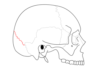
The lambdoid suture is a dense, fibrous connective tissue joint on the posterior aspect of the skull that connects the parietal bones with the occipital bone. It is continuous with the occipitomastoid suture.

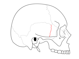
The coronal suture is a dense, fibrous connective tissue joint that separates the two parietal bones from the frontal bone of the skull.

The asterion is a meeting point between three sutures between bones of the skull. It is an important surgical landmark.

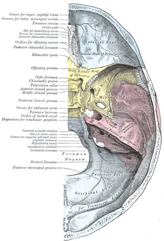
The middle cranial fossa is formed by the sphenoid bones, and the temporal bones. It lodges the temporal lobes, and the pituitary gland. It is deeper than the anterior cranial fossa, is narrow medially and widens laterally to the sides of the skull. It is separated from the posterior cranial fossa by the clivus and the petrous crest.
The frontoethmoidal suture is the suture between the ethmoid bone and the frontal bone.

The sphenozygomatic suture is the cranial suture between the sphenoid bone and the zygomatic bone.

The sphenoparietal suture is the cranial suture between the sphenoid bone and the parietal bone. It is one of the sutures that comprises the pterion.

The sphenofrontal suture is the cranial suture between the sphenoid bone and the frontal bone.

The zygomaticofrontal suture is the cranial suture between the zygomatic bone and the frontal bone. The suture can be palpated just lateral to the eye.

The zygomaticotemporal suture is the cranial suture between the zygomatic bone and the temporal bone. This is part of the zygomatic arch. Movement at the suture decreases with development during aging. It has a complex internal structure.
The sphenoethmoidal suture is the cranial suture between the sphenoid bone and the ethmoid bone.

The occipitomastoid suture or occipitotemporal suture is the cranial suture between the occipital bone and the mastoid portion of the temporal bone.
The sphenosquamosal suture is a cranial suture between the sphenoid bone and the squama of the temporal bone.

The pterion is the region where the frontal, parietal, temporal, and sphenoid bones join. It is located on the side of the skull, just behind the temple. It is also considered to be the weakest part of the skull, which makes it clinically significant, as if there is a fracture around the pterion it could be accompanied by an epidural hematoma.
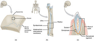
In anatomy, fibrous joints are joints connected by fibrous tissue, consisting mainly of collagen. These are fixed joints where bones are united by a layer of white fibrous tissue of varying thickness. In the skull, the joints between the bones are called sutures. Such immovable joints are also referred to as synarthroses.
In arthropod and vertebrate anatomy, the vertex is the highest point of the head.

The base of skull, also known as the cranial base or the cranial floor, is the most inferior area of the skull. It is composed of the endocranium and the lower parts of the calvaria.
This page is based on this
Wikipedia article Text is available under the
CC BY-SA 4.0 license; additional terms may apply.
Images, videos and audio are available under their respective licenses.

