The leg is the entire lower limb of the human body, including the foot, thigh or sometimes even the hip or buttock region. The major bones of the leg are the femur, tibia, and adjacent fibula. The thigh is between the hip and knee, while the calf (rear) and shin (front) are between the knee and foot.

The outer ear, external ear, or auris externa is the external part of the ear, which consists of the auricle and the ear canal. It gathers sound energy and focuses it on the eardrum.

The scapula, also known as the shoulder blade, is the bone that connects the humerus with the clavicle. Like their connected bones, the scapulae are paired, with each scapula on either side of the body being roughly a mirror image of the other. The name derives from the Classical Latin word for trowel or small shovel, which it was thought to resemble.
Articles related to anatomy include:

The auricle or auricula is the visible part of the ear that is outside the head. It is also called the pinna, a term that is used more in zoology.
Otoplasty is a procedure for correcting the deformities and defects of the pinna, whether these defects are congenital conditions or caused by trauma. Otoplastic surgeons may reshape, move, or augment the cartilaginous support framework of the pinna to correct these defects.
The helix is the prominent rim of the auricle. Where the helix turns downwards posteriorly, a small tubercle is sometimes seen, namely the auricular tubercle of Darwin.
The tragus is a small pointed eminence of the external ear, situated in front of the concha, and projecting backward over the meatus. It also is the name of hair growing at the entrance of the ear. Its name comes the Ancient Greek tragos, meaning 'goat', and is descriptive of its general covering on its under surface with a tuft of hair, resembling a goat's beard. The nearby antitragus projects forwards and upwards.

The abdominal external oblique muscle is the largest and outermost of the three flat abdominal muscles of the lateral anterior abdomen.

In human anatomy, the superficial temporal artery is a major artery of the head. It arises from the external carotid artery when it splits into the superficial temporal artery and maxillary artery.

The antihelix (anthelix) is a part of the visible ear; the pinna. The antihelix is a curved prominence of cartilage parallel with and in front of the helix on the pinna.
The antitragus is a feature of mammalian ear anatomy.

The antitragicus is an intrinsic muscle of the outer ear.

The tragicus, also called the tragus muscle or Valsalva muscle, is an intrinsic muscle of the outer ear.
The oblique muscle of auricle is an intrinsic muscle of the outer ear.
The transverse muscle of auricle is an intrinsic muscle of the outer ear.
The helicis major is an intrinsic muscle of the outer ear.

The anterior auricular branches of the superficial temporal artery are distributed to the anterior portion of the auricula, the lobule, and part of the external meatus, anastomosing with the posterior auricular. They supply the external acoustic meatus and the visible part of the ear.

The following outline is provided as an overview of and topical guide to human anatomy:

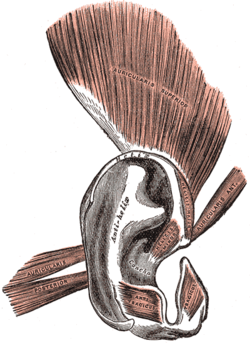
The anterior auricular muscle, the smallest of the three auricular muscles, is thin and fan-shaped, and its fibers are pale and indistinct. It arises from the lateral edge of the epicranial aponeurosis, and its fibers converge to be inserted into a projection on the front of the helix.
 Anatomy of human ear
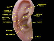 External ear. Right auricle.Lateral view.
 External ear. Right auricle.Lateral view.
 External ear. Right auricle.Lateral view.