This article is missing information about the situation in other tetrapods, cf Carpal bone#Other animals.(March 2025) |
| Lunate bone | |
|---|---|
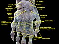
 Left hand anterior view (palmar view). Lunate bone shown in red. | |
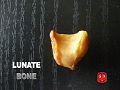
 The left lunate bone | |
| Details | |
| Articulations | Radius proximally capitate and hamate distally scaphoid laterally triangular medially triangular fibrocartilage [1] |
| Identifiers | |
| Latin | os lunatum |
| MeSH | D012667 |
| TA98 | A02.4.08.005 |
| TA2 | 1252 |
| FMA | 23712 |
| Anatomical terms of bone | |
The lunate bone (semilunar bone) is a carpal bone in the human hand. It is distinguished by its deep concavity and crescentic outline. It is situated in the center of the proximal row carpal bones, which lie between the ulna and radius and the hand. The lunate carpal bone is situated between the lateral scaphoid bone and medial triquetral bone.