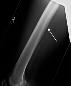
The humerus is a long bone in the arm that runs from the shoulder to the elbow. It connects the scapula and the two bones of the lower arm, the radius and ulna, and consists of three sections. The humeral upper extremity consists of a rounded head, a narrow neck, and two short processes. The body is cylindrical in its upper portion, and more prismatic below. The lower extremity consists of 2 epicondyles, 2 processes, and 3 fossae. As well as its true anatomical neck, the constriction below the greater and lesser tubercles of the humerus is referred to as its surgical neck due to its tendency to fracture, thus often becoming the focus of surgeons.

The sacrum, in human anatomy, is a large, triangular bone at the base of the spine that forms by the fusing of the sacral vertebrae (S1–S5) between ages 18 and 30.

The tibia, also known as the shinbone or shankbone, is the larger, stronger, and anterior (frontal) of the two bones in the leg below the knee in vertebrates ; it connects the knee with the ankle. The tibia is found on the medial side of the leg next to the fibula and closer to the median plane. The tibia is connected to the fibula by the interosseous membrane of leg, forming a type of fibrous joint called a syndesmosis with very little movement. The tibia is named for the flute tibia. It is the second largest bone in the human body, after the femur. The leg bones are the strongest long bones as they support the rest of the body.

In anatomy, the palatine bones are two irregular bones of the facial skeleton in many animal species, located above the uvula in the throat. Together with the maxilla, they comprise the hard palate.

The obturator foramen is the large, bilaterally paired opening of the bony pelvis. It is formed by the pubis and ischium. It is mostly closed by the obturator membrane except for a small opening, the obturator canal, through which the obturator nerve and vessels pass.
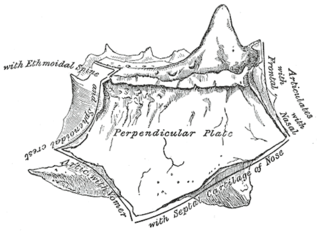
The perpendicular plate of the ethmoid bone is a thin, flattened lamina, polygonal in form, which descends from the under surface of the cribriform plate, and assists in forming the septum of the nose; it is generally deflected a little to one or other side. The anterior border articulates with the spine of the frontal bone and the crest of the nasal bones.

The vertebral vein is formed in the suboccipital triangle, from numerous small tributaries which spring from the internal vertebral venous plexuses and issue from the vertebral canal above the posterior arch of the atlas.

The optic foramen is the opening to the optic canal. The canal is located in the sphenoid bone; it is bounded medially by the body of the sphenoid and laterally by the lesser wing of the sphenoid.

The greater palatine foramen is, along with the lesser palatine foramen, one of two foramina (holes) in each of the left and right palatine bones which form the posterior roof of the human mouth, known as the palate. It is sometimes known as the major palatine foramen.

The posterior superior alveolar artery is a branch of the maxillary artery. It is one of two or three superior alveolar arteries. It provides arterial supply to the molar and premolar teeth, maxillary sinus and adjacent bone, and the gingiva.
In human anatomy, the body of femur is the almost cylindrical, long part of the femur. It is a little broader above than in the center, broadest and somewhat flattened from before backward below. It is slightly arched, so as to be convex in front, and concave behind, where it is strengthened by a prominent longitudinal ridge, the linea aspera.

The posterior superior alveolar nerves (also posterior superior dental nerves or posterior superior alveolar branches) are sensory branches of the maxillary nerve (CN V2). They arise within the pterygopalatine fossa as a single trunk. They run on or in the maxilla. They provide sensory innervation to the upper molar teeth and adjacent gum, and the maxillary sinus.

The zygomatico-orbital foramina are two canals in the skull, that allow nerves to pass through. The orifices are seen on the orbital process of the zygomatic bone.

The horizontal plate of palatine bone is a quadrilateral part of the palatine bone, and has two surfaces and four borders.
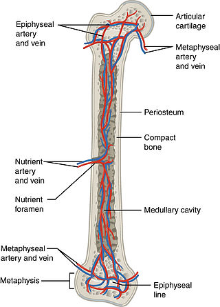
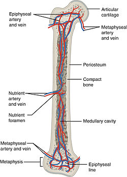
The nutrient artery, usually accompanied by one or two nutrient veins, enters the bone through the nutrient foramen, runs obliquely through the cortex, sends branches upward and downward to the bone marrow, which ramify in the endosteum–the vascular membrane lining the medullary cavity–and give twigs to the adjoining canals. Nutrient arteries are the most apparent blood vessels of the bones.

The femoral neck is a flattened pyramidal process of bone, connecting the femoral head with the femoral shaft, and forming with the latter a wide angle opening medialward.
The lumbar enlargement is a widened area of the spinal cord that gives attachment to the nerves which supply the lower limbs.
The cervical enlargement corresponds with the attachments of the large nerves which supply the upper limbs.

The following outline is provided as an overview of and topical guide to human anatomy:

Each vertebra is an irregular bone with a complex structure composed of bone and some hyaline cartilage, that make up the vertebral column or spine, of vertebrates. The proportions of the vertebrae differ according to their spinal segment and the particular species.