
In vertebrate anatomy, ribs are the long curved bones which form the rib cage, part of the axial skeleton. In most tetrapods, ribs surround the chest, enabling the lungs to expand and thus facilitate breathing by expanding the chest cavity. They serve to protect the lungs, heart, and other internal organs of the thorax. In some animals, especially snakes, ribs may provide support and protection for the entire body.
The rib cage is an endoskeletal enclosure in the thorax of most vertebrate animals that comprises the ribs, vertebral column and sternum, which protects vital organs such as the heart, lungs and great vessels. The circumferential enclosure formed by left and right rib cages, together known as the thoracic cage, is a semi-rigid bony and cartilaginous structure which surrounds the thoracic cavity and supports the shoulder girdles to form the core part of the axial skeleton.

The left and right brachiocephalic veins are major veins in the upper chest, formed by the union of the ipsilateral internal jugular vein and subclavian vein behind the sternoclavicular joint. The left brachiocephalic vein is more than twice the length of the right brachiocephalic vein.

In human anatomy, the thoracic duct is the larger of the two lymph ducts of the lymphatic system. The thoracic duct usually begins from the upper aspect of the cisterna chyli, passing out of the abdomen through the aortic hiatus into first the posterior mediastinum and then the superior mediastinum, extending as high up as the root of the neck before descending to drain into the systemic (blood) circulation at the venous angle.

The azygos vein is a vein running up the right side of the thoracic vertebral column draining itself towards the superior vena cava. It connects the systems of superior vena cava and inferior vena cava and can provide an alternative path for blood to the right atrium when either of the venae cavae is blocked.
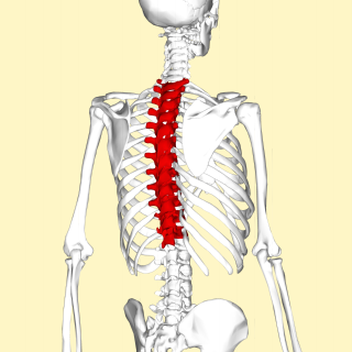
In vertebrates, thoracic vertebrae compose the middle segment of the vertebral column, between the cervical vertebrae and the lumbar vertebrae. In humans, there are twelve thoracic vertebrae and they are intermediate in size between the cervical and lumbar vertebrae; they increase in size going towards the lumbar vertebrae, with the lower ones being much larger than the upper. They are distinguished by the presence of facets on the sides of the bodies for articulation with the heads of the ribs, as well as facets on the transverse processes of all, except the eleventh and twelfth, for articulation with the tubercles of the ribs. By convention, the human thoracic vertebrae are numbered T1–T12, with the first one (T1) located closest to the skull and the others going down the spine toward the lumbar region.

The mediastinum is the central compartment of the thoracic cavity. Surrounded by loose connective tissue, it is an undelineated region that contains a group of structures within the thorax, namely the heart and its vessels, the esophagus, the trachea, the phrenic and cardiac nerves, the thoracic duct, the thymus and the lymph nodes of the central chest.

The transverse plane is an anatomical plane that divides the body into superior and inferior sections. It is perpendicular to the coronal and sagittal planes.
The superior thoracic aperture, also known as the thoracic outlet, or thoracic inlet refers to the opening at the top of the thoracic cavity. It is also clinically referred to as the thoracic outlet, in the case of thoracic outlet syndrome. A lower thoracic opening is the inferior thoracic aperture.

The internal intercostal muscles are a group of skeletal muscles located between the ribs. They are eleven in number on either side. They commence anteriorly at the sternum, in the intercostal spaces between the cartilages of the true ribs, and at the anterior extremities of the cartilages of the false ribs, and extend backward as far as the angles of the ribs, hence they are continued to the vertebral column by thin aponeuroses, the posterior intercostal membranes. They pull the sternum and ribs upward and inward.

The aortic arch, arch of the aorta, or transverse aortic arch is the part of the aorta between the ascending and descending aorta. The arch travels backward, so that it ultimately runs to the left of the trachea.

The hemiazygos vein is a vein running superiorly in the lower thoracic region, just to the left side of the vertebral column.

The sternoclavicular joint or sternoclavicular articulation is a synovial saddle joint between the manubrium of the sternum, and the clavicle, and the first costal cartilage. The joint possesses a joint capsule, and an articular disc, and is reinforced by multiple ligaments.

In human anatomy, the esophageal hiatus is an opening in the diaphragm through which the esophagus and the vagus nerve pass.
The xiphisternal joint is a location near the bottom of the sternum, where the body of the sternum and the xiphoid process meet. It is structurally classified as a synchondrosis, and functionally classified as a synarthrosis. The joint usually ossifies by the fourth decade of life, forming a synostosis.

The following outline is provided as an overview of and topical guide to human anatomy:

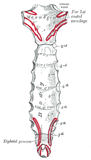
The sternum or breastbone is a long flat bone located in the central part of the chest. It connects to the ribs via cartilage and forms the front of the rib cage, thus helping to protect the heart, lungs, and major blood vessels from injury. Shaped roughly like a necktie, it is one of the largest and longest flat bones of the body. Its three regions are the manubrium, the body, and the xiphoid process. The word sternum originates from Ancient Greek στέρνον (stérnon) 'chest'.

Each vertebra is an irregular bone with a complex structure composed of bone and some hyaline cartilage, that make up the vertebral column or spine, of vertebrates. The proportions of the vertebrae differ according to their spinal segment and the particular species.

The pulmonary pleurae are the two opposing layers of serous membrane overlying the lungs and the inside of the surrounding chest walls.

