In vertebrate anatomy, ribs are the long curved bones which form the rib cage, part of the axial skeleton. In most tetrapods, ribs surround the thoracic cavity, enabling the lungs to expand and thus facilitate breathing by expanding the thoracic cavity. They serve to protect the lungs, heart, and other vital organs of the thorax. In some animals, especially snakes, ribs may provide support and protection for the entire body.

The rib cage or thoracic cage, is an endoskeletal enclosure in the thorax of most vertebrates that comprises the ribs, vertebral column and sternum, which protect the vital organs of the thoracic cavity, such as the heart, lungs and great vessels and support the shoulder girdle to form the core part of the axial skeleton.
The human skeleton is the internal framework of the human body. It is composed of around 270 bones at birth – this total decreases to around 206 bones by adulthood after some bones get fused together. The bone mass in the skeleton makes up about 14% of the total body weight and reaches maximum mass between the ages of 25 and 30. The human skeleton can be divided into the axial skeleton and the appendicular skeleton. The axial skeleton is formed by the vertebral column, the rib cage, the skull and other associated bones. The appendicular skeleton, which is attached to the axial skeleton, is formed by the shoulder girdle, the pelvic girdle and the bones of the upper and lower limbs.

The clavicle, collarbone, or keybone is a slender, S-shaped long bone approximately 6 inches (15 cm) long that serves as a strut between the shoulder blade and the sternum (breastbone). There are two clavicles, one on the left and one on the right. The clavicle is the only long bone in the body that lies horizontally. Together with the shoulder blade, it makes up the shoulder girdle. It is a palpable bone and, in people who have less fat in this region, the location of the bone is clearly visible. It receives its name from Latin clavicula 'little key' because the bone rotates along its axis like a key when the shoulder is abducted. The clavicle is the most commonly fractured bone. It can easily be fractured by impacts to the shoulder from the force of falling on outstretched arms or by a direct hit.

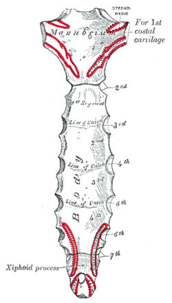
The xiphoid process, also referred to as the ensiform process, xiphisternum, or metasternum, constitutes a small cartilaginous process (extension) located in the inferior segment of the sternum, typically ossified in adult humans. Both the Greek-derived term xiphoid and its Latin equivalent, ensiform, connote a "swordlike" or "sword-shaped" morphology.

The left and right brachiocephalic veins are major veins in the upper chest, formed by the union of the ipsilateral internal jugular vein and subclavian vein behind the sternoclavicular joint. The left brachiocephalic vein is more than twice the length of the right brachiocephalic vein.

The axial skeleton is the part of the skeleton that consists of the bones of the head and trunk of a vertebrate. In the human skeleton, it consists of 80 bones and is composed of six parts; the skull, also the ossicles of the middle ear, the hyoid bone, the rib cage, sternum and the vertebral column. The axial skeleton together with the appendicular skeleton form the complete skeleton. Another definition of axial skeleton is the bones including the vertebrae, sacrum, coccyx, skull, ribs, and sternum.

The pectoralis major is a thick, fan-shaped or triangular convergent muscle of the human chest. It makes up the bulk of the chest muscles and lies under the breast. Beneath the pectoralis major is the pectoralis minor muscle.

The sternal angle is the projecting angle formed between the manubrium and body of a sternum at their junction at the manubriosternal joint.

The sternohyoid muscle is a bilaterally paired, long, thin, narrow strap muscle of the anterior neck. It is one of the infrahyoid muscles. It is innervated by the ansa cervicalis. It acts to depress the hyoid bone.

The transverse cervical nerve is a cutaneous (sensory) nerve of the cervical plexus that arises from the second and third cervical spinal nerves (C2-C3). It curves around the posterior border of the sternocleidomastoideus muscle, then pierces the fascia of the neck before dividing into two branches. It provides sensory innervation to the front of the neck.

The sternoclavicular joint or sternoclavicular articulation is a synovial saddle joint between the manubrium of the sternum, and the clavicle, and the first costal cartilage. The joint possesses a joint capsule, and an articular disc, and is reinforced by multiple ligaments.

The intertrochanteric line is a line upon the anterior aspect of the proximal end of the femur, extending between the lesser trochanter and the greater trochanter. It is a rough, variable ridge.

The sternocostal joints, also known as sternochondral joints or costosternal articulations, are synovial plane joints of the costal cartilages of the true ribs with the sternum. The only exception is the first rib, which has a synchondrosis joint since the cartilage is directly united with the sternum. The sternocostal joints are important for thoracic wall mobility.
The anterior sternoclavicular ligament is a broad band of fibers attached to the clavicle above, and to the manubrium below. The ligament overlies the anterior (front) surface of sternoclavicular joint.

The sternalismuscle is an anatomical variation that lies in front of the sternal end of the pectoralis major parallel to the margin of the sternum. The sternalis muscle may be a variation of the pectoralis major or of the rectus abdominis.
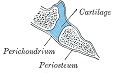
A synchondrosis is a type of cartilaginous joint where hyaline cartilage completely joins together two bones. Synchondroses are different from symphyses which are formed of fibrocartilage. Synchondroses are immovable joints and are thus referred to as synarthroses.are all synchondroses synarthrotic/immovable

Cartilaginous joints are connected entirely by cartilage. Cartilaginous joints allow more movement between bones than a fibrous joint but less than the highly mobile synovial joint. Cartilaginous joints also forms the growth regions of immature long bones and the intervertebral discs of the spinal column.

The sternum or breastbone is a long flat bone located in the central part of the chest. It connects to the ribs via cartilage and forms the front of the rib cage, thus helping to protect the heart, lungs, and major blood vessels from injury. Shaped roughly like a necktie, it is one of the largest and longest flat bones of the body. Its three regions are the manubrium, the body, and the xiphoid process. The word sternum originates from Ancient Greek στέρνον (stérnon) 'chest'.

Many anatomical terms descriptive of bone are defined in anatomical terminology, and are often derived from Greek and Latin. Bone in the human body is categorized into long bone, short bone, flat bone, irregular bone and sesamoid bone.