The outer ear, external ear, or auris externa is the external part of the ear, which consists of the auricle and the ear canal. It gathers sound energy and focuses it on the eardrum.

The auricle or auricula is the visible part of the ear that is outside the head. It is also called the pinna, a term that is used more in zoology.

The cervical plexus is a nerve plexus of the anterior rami of the first four cervical spinal nerves C1-C4. The cervical plexus provides motor innervation to some muscles of the neck, and the diaphragm; it provides sensory innervation to parts of the head, neck, and chest.
Otoplasty is a procedure for correcting the deformities and defects of the pinna, whether these defects are congenital conditions or caused by trauma. Otoplastic surgeons may reshape, move, or augment the cartilaginous support framework of the pinna to correct these defects.
The helix is the prominent rim of the auricle. Where the helix turns downwards posteriorly, a small tubercle is sometimes seen, namely the auricular tubercle of Darwin.

The transverse muscle of tongue is an intrinsic muscle of the tongue. It consists of fibers which arise from the median fibrous septum. It passes laterally to insert into the submucous fibrous tissue at the sides of the tongue. It is innervated by the hypoglossal nerve. Its contraction elongates and narrows the tongue.

The occipital artery is a branch of the external carotid artery that provides arterial supply to the back of the scalp, sternocleidomastoid muscles, and deep muscles of the back and neck.

The posterior auricular vein is a vein of the head. It begins from a plexus with the occipital vein and the superficial temporal vein, descends behind the auricle, and drains into the external jugular vein.
The antitragus is a feature of mammalian ear anatomy.

The posterior auricular nerve is a nerve of the head. It is a branch of the facial nerve. It communicates with branches from the vagus nerve, the great auricular nerve, and the lesser occipital nerve. Its auricular branch supplies the posterior auricular muscle, the intrinsic muscles of the auricle, and gives sensation to the auricle. Its occipital branch supplies the occipitalis muscle.

The antitragicus is an intrinsic muscle of the outer ear.

The tragicus, also called the tragus muscle or Valsalva muscle, is an intrinsic muscle of the outer ear.
The oblique muscle of auricle is an intrinsic muscle of the outer ear.
The Helicis minor is a small skeletal muscle. The helicis minor is an intrinsic muscle of the outer ear. The muscle runs obliques and covers the helical crus, part of the helix located just above the tragus.
The helicis major is an intrinsic muscle of the outer ear.

The following outline is provided as an overview of and topical guide to human anatomy:
The posterior auricular muscle is a muscle behind the auricle of the outer ear. It arises from the mastoid part of the temporal bone, and inserts into the lower part of the cranial surface of the auricle of the outer ear. It draws the auricle backwards, usually a very slight effect.

The superior auricular muscle is a muscle above the auricle of the outer ear. It originates from the epicranial aponeurosis, and inserts into the upper part of the medial surface of the auricle. It draws the auricle upwards.

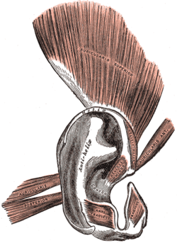
The anterior auricular muscle, the smallest of the three auricular muscles, is thin and fan-shaped, and its fibers are pale and indistinct. It arises from the lateral edge of the epicranial aponeurosis, and its fibers converge to be inserted into a projection on the front of the helix.
The oculo-auricular phenomenon, first described by Kinnier Wilson in 1908, is the phenomenon of an extreme lateral gaze inducing a slight but perceptible backwards movement of the upper part of the pinna. It is a muscle synergy involving the Abducens innervated lateral rectus muscle, an external muscle of the eye, and the facial innervated posterior auricular muscle, an external muscle of the ear. Wilson's phenomenon had attracted attention at the time because of his renown and for its implications regarding Darwin's theory of natural selection. According to, "In patients with brainstem disease abnormal transverse auricular muscle coactivation is characterized by absence of activity in one or both ear muscles during lateral gaze in either or both directions."
 Anatomy of human ear