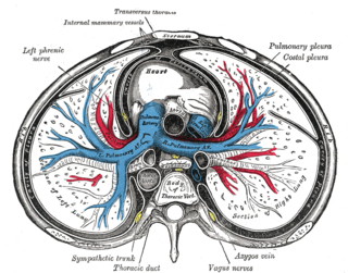
The pleural cavity is the thin fluid-filled space between the two pulmonary pleurae of each lung. A pleura is a serous membrane which folds back onto itself to form a two-layered membranous pleural sac. The outer pleura is attached to the chest wall, but is separated from it by the endothoracic fascia. The inner pleura covers the lungs and adjoining structures, including blood vessels, bronchi and nerves. The pleural cavity can be viewed as a potential space because the two pleurae adhere to each other under all normal conditions. Parietal pleura projects up to 2.5 cm above the junction of the middle and medial third of the clavicle
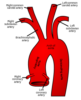
In human anatomy, the subclavian arteries are paired major arteries of the upper thorax, below the clavicle. They receive blood from the aortic arch. The left subclavian artery supplies blood to the left arm and the right subclavian artery supplies blood to the right arm, with some branches supplying the head and thorax. On the left side of the body, the subclavian comes directly off the aortic arch, while on the right side it arises from the relatively short brachiocephalic artery when it bifurcates into the subclavian and the right common carotid artery.
A fascia is a band or sheet of connective tissue, primarily collagen, beneath the skin that attaches, stabilizes, encloses, and separates muscles and other internal organs. Fascia is classified by layer, as superficial fascia, deep fascia, and visceral or parietal fascia, or by its function and anatomical location.

Hubert von Luschka, born Hubert Luschka, was a German anatomist. He lent his name to several structures, including the foramina of Luschka, Luschka's crypts, Luschka's joints, and Ducts of Luschka. His name is also associated with Luschka's law, a anatomical rule concerning location of the ureters.

In anatomy, serous membrane is a smooth tissue membrane consisting of two layers of mesothelium, which secrete serous fluid. The inner layer that covers organs (viscera) in body cavities is called the visceral membrane. A second layer of epithelial cells of the serous membrane, called the parietal layer, lines the body wall. Between the two layers is a potential space, mostly empty except for a few milliliters of lubricating serous fluid that is secreted by the two serous membranes.

The stellate ganglion is a sympathetic ganglion formed by the fusion of the inferior cervical ganglion and the first thoracic ganglion, which exists in 80% of cases. Sometimes the second and the third thoracic ganglia are included in this fusion. Stellate ganglion is relatively big compared to much smaller thoracic, lumbar and sacral ganglia and it is polygonal in shape. Stellate ganglion is located at the level of C7, anterior to the transverse process of C7 and the neck of the first rib, superior to the cervical pleura and just below the subclavian artery. It is superiorly covered by the prevertebral lamina of the cervical fascia and anteriorly in relation with common carotid artery, subclavian artery and the beginning of vertebral artery which sometimes leaves a groove at the apex of this ganglion. Relations of the apex of the stellate ganglion: • covered by the endothoracic fascia and parietal pleura • right stellate ganglion is in relation with right brachiocephalic vein anteriorly • right stellate ganglion is in relation with sternal part of subclavian artery anteriorly • laterally: first intercostal artery • medially: longus colli muscle

The transversus thoracis muscle lies internal to the thoracic cage, anteriorly. It is a thin plane of muscular and tendinous fibers, situated upon the inner surface of the front wall of the chest. It is in the same layer as the subcostal muscles and the innermost intercostal muscles.
In anatomy, the left and right common carotid arteries (carotids) are arteries that supply the head and neck with oxygenated blood; they divide in the neck to form the external and internal carotid arteries.

The intercostal nerves are part of the somatic nervous system, and arise from the anterior rami of the thoracic spinal nerves from T1 to T11. The intercostal nerves are distributed chiefly to the thoracic pleura and abdominal peritoneum and differ from the anterior rami of the other spinal nerves in that each pursues an independent course without plexus formation.
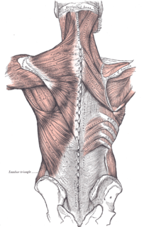
The thoracolumbar fascia is a deep investing membrane throughout most of the posterior thorax and abdomen although it is a thin fibrous lamina in the thoracic region. Above, it is continuous with a similar investing layer on the back of the neck—the nuchal fascia.

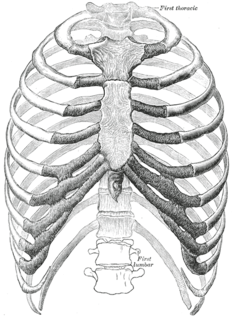
The intercostal space (ICS) is the anatomic space between two ribs. Since there are 12 ribs on each side, there are 11 intercostal spaces, each numbered for the rib superior to it.

The membranous layer of the superficial fascia of the perineum is the deeper layer of the superficial perineal fascia. It is thin, aponeurotic in structure, and of considerable strength, serving to bind down the muscles of the root of the penis. Colles' fascia emerges from the perineal membrane, which divides the base of the penis from the prostate. Colles' fascia emerges from the inferior side of the perineal membrane and continues along the ventral (inferior) penis without covering the scrotum. It separates the skin and subcutaneous fat from the superficial perineal pouch.

The costocervical trunk arises from the upper and back part of the second part of subclavian artery, behind the scalenus anterior on the right side, and medial to that muscle on the left side.

The thoracic wall or chest wall is the boundary of the thoracic cavity.
The pectoral fascia is a thin lamina, covering the surface of the pectoralis major, and sending numerous prolongations between its fasciculi: it is attached, in the middle line, to the front of the sternum; above, to the clavicle; laterally and below it is continuous with the fascia of the shoulder, axilla, and thorax.
The suprapleural membrane, eponymously known as Sibson's fascia, is a structure described in human anatomy.

The perineal membrane is an anatomical term for a fibrous membrane in the perineum. The term "inferior fascia of urogenital diaphragm", used in older texts, is considered equivalent to the perineal membrane.

The external spermatic fascia is a thin membrane, prolonged downward around the surface of the spermatic cord and testis. It is separated from the dartos tunic by loose areolar tissue. It is occasionally referred to as 'Le Fascia de Webster' after an anatomist who once described it.

The renal fascia or Gerota's fascia is a layer of connective tissue encapsulating the kidneys and the adrenal glands. The renal fascia separates the adipose capsule of kidney from the overlying pararenal fat. The deeper layers below the renal fascia are, in order, the adipose capsule, the renal capsule and finally the parenchyma of the renal cortex. The spaces about the kidney are typically divided into three compartments: the perinephric space and the anterior and posterior pararenal spaces.