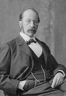
The theory of recapitulation, also called the biogenetic law or embryological parallelism—often expressed using Ernst Haeckel's phrase "ontogeny recapitulates phylogeny"—is an historical hypothesis that the development of the embryo of an animal, from fertilization to gestation or hatching (ontogeny), goes through stages resembling or representing successive adult stages in the evolution of the animal's remote ancestors (phylogeny). It was formulated in the 1820s by Étienne Serres based on the work of Johann Friedrich Meckel, after whom it is also known as Meckel–Serres law.
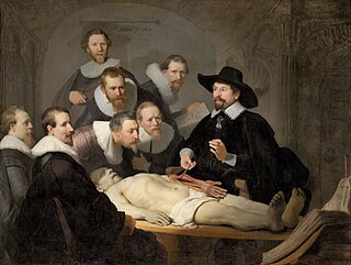
An autopsy is a surgical procedure that consists of a thorough examination of a corpse by dissection to determine the cause, mode, and manner of death; or the exam may be performed to evaluate any disease or injury that may be present for research or educational purposes. The term necropsy is generally used for non-human animals.

Dissection is the dismembering of the body of a deceased animal or plant to study its anatomical structure. Autopsy is used in pathology and forensic medicine to determine the cause of death in humans. Less extensive dissection of plants and smaller animals preserved in a formaldehyde solution is typically carried out or demonstrated in biology and natural science classes in middle school and high school, while extensive dissections of cadavers of adults and children, both fresh and preserved are carried out by medical students in medical schools as a part of the teaching in subjects such as anatomy, pathology and forensic medicine. Consequently, dissection is typically conducted in a morgue or in an anatomy lab.

Ernst Heinrich Weber was a German physician who is considered one of the founders of experimental psychology. He was an influential and important figure in the areas of physiology and psychology during his lifetime and beyond. His studies on sensation and touch, along with his emphasis on good experimental techniques led to new directions and areas of study for future psychologists, physiologists, and anatomists.

Jan Evangelista Purkyně was a Czech anatomist and physiologist. In 1839, he coined the term "protoplasma" for the fluid substance of a cell. He was one of the best known scientists of his time. Such was his fame that when people from outside Europe wrote letters to him, all that they needed to put as the address was "Purkyně, Europe".
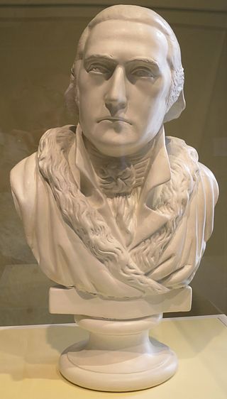
Philip Syng Physick was an American physician and professor born in Philadelphia. He was the first professor of surgery and later of anatomy at the University of Pennsylvania medical school from 1805 to 1831 during which time he was a highly influential teacher. Physick invented a number of surgical devices and techniques including the stomach tube and absorbable sutures. He has been called the "Father of American Surgery."

Albert von Kölliker was a Swiss anatomist, physiologist, and histologist.
A microtome is a cutting tool used to produce extremely thin slices of material known as sections, with the process being termed microsectioning. Important in science, microtomes are used in microscopy for the preparation of samples for observation under transmitted light or electron radiation.

Wilhelm Roux was a German zoologist and pioneer of experimental embryology.
Bone decalcification is the softening of bones due to the removal of calcium ions, and can be performed as a histological technique to study bones and extract DNA. This process also occurs naturally during bone development and growth, and when uninhibited, can cause diseases such as osteomalacia.

In amniote embryonic development, the epiblast is one of two distinct cell layers arising from the inner cell mass in the mammalian blastocyst, or from the blastula in reptiles and birds, the other layer is the hypoblast. It drives the embryo proper through its differentiation into the three primary germ layers, ectoderm, mesoderm and endoderm, during gastrulation. The amniotic ectoderm and extraembryonic mesoderm also originate from the epiblast.

Karl Bogislaus Reichert was a German anatomist, embryologist and histologist.

Christian Wilhelm Braune was a German anatomist. He is known for his excellent lithographs of cross-sections of the human body, and his pioneer work in biomechanics. He also pioneered the use of frozen cadavers for anatomical investigations.

Carl Rabl was an Austrian anatomist. His most notable achievement was on the structural consistency of chromosomes during the cell cycle. In 1885 he published that chromosomes do not lose their identity, even though they are no longer visible through the microscope.
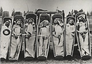
A cadaver or corpse is a dead human body. Cadavers are used by medical students, physicians and other scientists to study anatomy, identify disease sites, determine causes of death, and provide tissue to repair a defect in a living human being. Students in medical school study and dissect cadavers as a part of their education. Others who study cadavers include archaeologists and arts students. In addition, a cadaver may be used in the development and evaluation of surgical instruments.
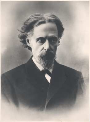
August Rauber was a German anatomist and embryologist born in Obermoschel in the Rhineland-Palatinate.

The rhombic lip is a posterior section of the developing metencephalon which can be recognized transiently within the vertebrate embryo. It extends posteriorly from the roof of the fourth ventricle to dorsal neuroepithelial cells. The rhombic lip can be divided into eight structural units based on rhombomeres 1-8 (r1-r8), which can be recognized at early stages of hindbrain development. Producing granule cells and five brainstem nuclei, the rhombic lip plays an important role in developing a complex cerebellar neural system.

Franklin Paine Mall was an American anatomist and pathologist known for his research and literature in the fields of anatomy and embryology. Mall was granted a fellowship for the Department of Pathology at the Johns Hopkins University and after positions at other universities, later returned to be the head of the first Anatomy Department at the Johns Hopkins School of Medicine. There, he reformed the field of anatomy and its educational curriculum. Mall was the founder and the first chief of the Department of Embryology at the Carnegie Institution for Science. He later donated his collection of human embryos that he started as a postgraduate student to the Carnegie Institution for Science. He was an elected member of the American Academy of Arts and Sciences, the American Philosophical Society, and the United States National Academy of Sciences.
The necrobiome has been defined as the community of species associated with decaying corpse remains. The process of decomposition is complex. Microbes decompose cadavers, but other organisms including fungi, nematodes, insects, and larger scavenger animals also contribute. Once the immune system is no longer active, microbes colonizing the intestines and lungs decompose their respective tissues and then travel throughout the body via the blood and lymphatic systems to break down other tissue and bone. During this process, gases are released as a by-product and accumulate, causing bloating. Eventually, the gases seep through the body's wounds and natural openings, providing a way for some microbes to exit from the inside of the cadaver and inhabit the outside. The microbial communities colonizing the internal organs of a cadaver are referred to as the thanatomicrobiome. The region outside of the cadaver that is exposed to the external environment is referred to as the epinecrotic portion of the necrobiome, and is especially important when determining the time and location of death for an individual. Different microbes play specific roles during each stage of the decomposition process. The microbes that will colonize the cadaver and the rate of their activity are determined by the cadaver itself and the cadaver's surrounding environmental conditions.
George Linius Streeter PAAA NAS APS HFRSE (1873–1948) was a 20th-century American anatomist and world leading embryologist. He was Director of the Carnegie Institution of Washington from 1917 to 1940.
