Albert von Kölliker

Last updated • 7 min readFrom Wikipedia, The Free Encyclopedia

Rudolf Albert von Kölliker
Kolliker Rudolph Albert von 1818-1902.jpg
Born
Rudolf Albert Kölliker

(1817-07-06)6 July 1817
Died2 November 1905(1905-11-02) (aged 88)
NationalitySwiss
Alma mater University of Zurich
University of Bonn
University of Berlin
Known forContributions to zoology
Awards Copley Medal (1897)
Linnean Medal (1902)
Scientific career
Fields Anatomy, physiology
Doctoral advisor Johannes Peter Müller
Friedrich Gustav Jakob Henle

Albert von Kölliker (born Rudolf Albert Kölliker; 6 July 1817 – 2 November 1905) was a Swiss anatomist, physiologist, and histologist.

Contents

Biography

Albert Kölliker was born in Zürich, Switzerland. His early education was carried on in Zürich, and he entered the university there in 1836. After two years, however, he moved to the University of Bonn, and later to that of Berlin, becoming a pupil of noted physiologists Johannes Peter Müller and of Friedrich Gustav Jakob Henle. He graduated in philosophy at Zürich in 1841, and in medicine at Heidelberg in 1842 The first academic post which he held was that of prosector of anatomy under Henle, but his tenure of this office was brief in 1844 he returned to University of Zurich to occupy a chair as professor extraordinary of physiology and comparative anatomy. His stay here was also brief; in 1847 the University of Würzburg, attracted by his rising fame, offered him the post of professor of physiology and of microscopical and comparative anatomy. He accepted the appointment, and at Würzburg he remained thenceforth, refusing all offers tempting him to leave the quiet academic life of the Bavarian town, where he died. [1]

Many of the numerous memoirs which he published, (including the very first paper he wrote) and which appeared in 1841, before he graduated, were on the structure of animals of the most varied kinds. Notable among these were his papers on the Medusae and allied creatures. His activity in this direction led him to make zoological excursions to the Mediterranean Sea and to the coasts of Scotland, as well as to undertake, conjointly with his friend Carl Theodor Ernst von Siebold, the editorship of the Zeitschrift für Wissenschaftliche Zoologie, which, founded in 1848, continued under his hands to be one of the most important zoological periodicals. [2]

His hand was one of the first to be x-rayed, by his friend Wilhelm Röntgen. [3]

X-ray of Kolliker's hand, made by Rontgen on 23 Jan 1896 X-ray by Wilhelm Rontgen of Albert von Kolliker's hand - 18960123-02.jpg
X-ray of Kölliker's hand, made by Röntgen on 23 Jan 1896

Works

Kölliker made contributions to the study of zoology. His earlier efforts were directed to the invertebrates, and his memoir on the development of cephalopods (which appeared in 1844) is considered a classical work. He soon passed on to the vertebrates, and studied the amphibians and mammalian embryos. He was among the first, if not the very first, to introduce into this branch of biological inquiry the newer microscopic technique – the methods of hardening, sectioning and staining. Much of the progress which embryology made during the middle and latter half of the 19th century are associated with his name.[ citation needed ] His Lectures on Development, published in 1861, at once became a standard work. [2]

But neither zoology nor embryology furnished Kölliker's chief claim to fame. If he did much for these branches of science, he did still more for histology, the knowledge of the minute structure of the animal tissues. Among his earlier results was the demonstration in 1847 that smooth or unstriated muscle is made up of distinct units, of nucleated muscle cells. In this work, he followed in the footsteps of his master Henle. A few years before this, there was doubt whether arteries had muscle in their walls – in addition, no solid histological basis as yet existed for those views as to the action of the nervous system on the circulation, which were soon to be put forward, and which had such a great influence on the progress of physiology. [2]

Kölliker's contributions to histology were widespread; smooth muscle, striated muscle, skin, bone, teeth, blood vessels and viscera were all investigated by Kölliker. The results at which he arrived were recorded partly in separate memoirs, partly in his great textbook on microscopical anatomy, which first saw the light in 1850. [2]

Albert L. Lehninger asserted that Kölliker was among the first to notice the arrangement of granules in the sarcoplasm of striated muscle over a period of years beginning around 1850. These granules were later called sarcosomes by Retzius in 1890. These sarcosomes have come to be known as the mitochondria-the power houses of the cell. In the words of Lehninger, "Kölliker should also be credited with the first separation of mitochondria from cell structure. In 1888 he teased these granules from insect muscle, in which they are very profuse, found them to swell in water, and showed them to possess a membrane."[ citation needed ]

In the case of almost every tissue, our present knowledge contains information first discovered by Kölliker – it is for his work on the nervous system that his name is most remembered. As early as 1845, while still at Zürich, he supplied a clear proof that nerve fibers are continuous with nerve cells, and laying the groundwork for all later research as to the actions of the central nervous system. [2]

From that time onward he continually laboured at the histology of the nervous system, and more especially at the difficult problems presented by the intricate patterns in which nerve fibers and neurons are woven together in the brain and spinal cord. From his early days a master of method, he saw at a glance the value of the new Golgi staining method for the investigation of the central nervous system. Kölliker contributed greatly to knowledge of the inner structure of the brain. [2] [4] In 1889, he reproduces the histological preparations of the father of neuroscience Ramón y Cajal and confirmed the theory of neuronism.

Honors

Kölliker was ennobled by prince regent Luitpold of Bavaria in 1897 and thus permitted to add the predicate "von" to his surname.[ citation needed ] He was made a member of the learned societies of many countries; in England, which he visited more than once, and where he became well known, the Royal Society made him a fellow in 1860, and in 1897 gave him its highest token of esteem, the Copley medal. [2]

A species of lizard, Hyalosaurus koellikeri , is named in his honor. [5]

Heterogenesis

In 1864 Kölliker revived Étienne Geoffroy Saint-Hilaire's theory that evolution proceeds by large steps (saltationism), under the name of heterogenesis. [7] Kölliker was a critic of Darwinism and rejected a universal common ancestor, instead he supported a theory of common descent along separate lines. [8] According to Alexander Vucinich the non-Darwinian evolution theory of Kölliker tied "organic transformism to three general ideas, all contrary to Darwin's view: the multiple origin of living forms, the internal causes of variation, and "sudden leaps" (heterogenesis) in the evolutionary process." [9] Kölliker claimed that heterogenesis functioned according to a general law of evolutionary progress, orthogenesis. [10]

Notes

  1. Foster 1911, p. 889.
  2. 1 2 3 4 5 6 7 Foster 1911, p. 890.
  3. "RÖNTGEN, Wilhelm Conrad (1845–1923). Ueber eine neue Art von Strahlen (Vorläufige Mittheilung). – Eine neue Art von Strahlen. II. Mittheilung. Offprints from: Sitzungsberichte der Würzburger Physik.-medic. Gesellschaft, 1895 [no. 9], and 1896, [nos. 1–2]. Würzburg: Verlag und Druck der Stahel'schen k. Hof.-und Universitäts- Buch- und Kunsthandlung, 1895–1896". www.christies.com. Retrieved 3 April 2019.
  4. Handbuch der Gewebelehre des Menschen, t. 2, Leipzig, 1896. (in German).
  5. Beolens, Bo; Watkins, Michael; Grayson, Michael (2011). The Eponym Dictionary of Reptiles. Baltimore: Johns Hopkins University Press. xiii + 296 pp. ISBN   978-1-4214-0135-5. ("Koelliker", p. 144).
  6. International Plant Names Index.  Koell.
  7. Wright, Sewall (1984). Evolution and the Genetics of Populations: Genetics and Biometric Foundations . Vol. 1. University of Chicago Press. p.  10. ISBN   978-0-226-91049-9.
  8. Di Gregorio, Mario A. (2005). From Here to Eternity: Ernst Haeckel and Scientific Faith. Vandenhoeck & Ruprecht. p. 303. ISBN   978-3-525-56972-6.
  9. Vucinich, Alexander (1988). Darwin in Russian Thought. University of California Press. p. 137. ISBN   978-0-520-06283-2.
  10. Glick, Thomas F. (1988). The Comparative Reception of Darwinism. University of Chicago Press. p. 99. ISBN   978-0-226-29977-8.

Related Research Articles

<span class="mw-page-title-main">Anatomy</span> Study of the structure of organisms and their parts

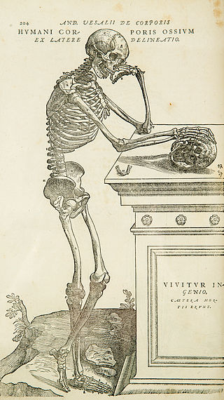
Anatomy is the branch of morphology concerned with the study of the internal structure of organisms and their parts. Anatomy is a branch of natural science that deals with the structural organization of living things. It is an old science, having its beginnings in prehistoric times. Anatomy is inherently tied to developmental biology, embryology, comparative anatomy, evolutionary biology, and phylogeny, as these are the processes by which anatomy is generated, both over immediate and long-term timescales. Anatomy and physiology, which study the structure and function of organisms and their parts respectively, make a natural pair of related disciplines, and are often studied together. Human anatomy is one of the essential basic sciences that are applied in medicine, and is often studied alongside physiology.

<span class="mw-page-title-main">Histology</span> Study of the microscopic anatomy of cells and tissues of plants and animals

Histology, also known as microscopic anatomy or microanatomy, is the branch of biology that studies the microscopic anatomy of biological tissues. Histology is the microscopic counterpart to gross anatomy, which looks at larger structures visible without a microscope. Although one may divide microscopic anatomy into organology, the study of organs, histology, the study of tissues, and cytology, the study of cells, modern usage places all of these topics under the field of histology. In medicine, histopathology is the branch of histology that includes the microscopic identification and study of diseased tissue. In the field of paleontology, the term paleohistology refers to the histology of fossil organisms.

<span class="mw-page-title-main">Friedrich Gustav Jakob Henle</span> German physician, pathologist, and anatomist (1809–1885)

Friedrich Gustav Jakob Henle was a German physician, pathologist, and anatomist. He is credited with the discovery of the loop of Henle in the kidney. His essay, "On Miasma and Contagia," was an early argument for the germ theory of disease. He was an important figure in the development of modern medicine.

<span class="mw-page-title-main">Carl Gegenbaur</span> German anatomist (1826–1903)

Carl Gegenbaur was a German anatomist and professor who demonstrated that the field of comparative anatomy offers important evidence supporting of the theory of evolution. As a professor of anatomy at the University of Jena (1855–1873) and at the University of Heidelberg (1873–1903), Carl Gegenbaur was a strong supporter of Charles Darwin's theory of organic evolution, having taught and worked, beginning in 1858, with Ernst Haeckel, eight years his junior.

<span class="mw-page-title-main">Ádám Politzer</span>

Ádám Politzer was a Hungarian and Austrian physician and one of the pioneers and founders of otology.

<span class="mw-page-title-main">Nissl body</span> Rough endoplasmic reticulum structure found in neurons

In cellular neuroscience, Nissl bodies are discrete granular structures in neurons that consist of rough endoplasmic reticulum, a collection of parallel, membrane-bound cisternae studded with ribosomes on the cytosolic surface of the membranes. Nissl bodies were named after Franz Nissl, a German neuropathologist who invented the staining method bearing his name. The term "Nissl bodies" generally refers to discrete clumps of rough endoplasmic reticulum and free ribosomes in nerve cells. Masses of rough endoplasmic reticulum also occur in some non-neuronal cells, where they are referred to as ergastoplasm, basophilic bodies, or chromophilic substance. While these organelles differ in some ways from Nissl bodies in neurons, large amounts of rough endoplasmic reticulum are generally linked to the copious production of proteins.

<span class="mw-page-title-main">Endoneurium</span> Connective tissue layer around myelinated nerve fibers in peripheral nervous system

The endoneurium is a layer of delicate connective tissue around the myelin sheath of each myelinated nerve fiber in the peripheral nervous system. Its component cells are called endoneurial cells. The endoneuria with their enclosed nerve fibers are bundled into groups called nerve fascicles, each fascicle within its own protective sheath called a perineurium. In sufficiently large nerves multiple fascicles, each with its blood supply and fatty tissue, may be bundled within yet another sheath, the epineurium.

<span class="mw-page-title-main">Franz Leydig</span> German zoologist and comparative anatomist

Franz von Leydig, also Franz Leydig, was a German zoologist and comparative anatomist.

Heinrich Müller was a German anatomist and professor at the University of Würzburg. He is best known for his work in comparative anatomy and his studies involving the eye.

<span class="mw-page-title-main">Vladimir Betz</span> Ukrainian anatomist and histologist

Vladimir Alexeyevich Betz, also Volodymyr Oleksiyovych Betz was a Russian and Ukrainian anatomist and histologist, served as a professor at the Saint Vladimir University of Kyiv.

Martin Heidenhain was a German anatomist born in Breslau. His father was physiologist Rudolf Heidenhain (1834-1937), and his mother, Fanny Volkmann, was the daughter of anatomist Alfred Wilhelm Volkmann (1800-1877).

<span class="mw-page-title-main">Eusebio Oehl</span> Italian histologist and physiologist

Eusebio Oehl was an Italian histologist and physiologist who was a native of Lodi.

<span class="mw-page-title-main">Victor von Ebner</span> Austrian anatomist and histologist

Anton Gilbert Victor von Ebner, Ritter von Rofenstein was an Austrian anatomist and histologist.

<span class="mw-page-title-main">Karl Joseph Eberth</span> German pathologist (1835–1926)

Karl Joseph Eberth was a German pathologist and bacteriologist who was a native of Würzburg.

<span class="mw-page-title-main">Mihály Lenhossék</span> Hungarian anatomist and histologist

Mihály Lenhossék, named often given as Michael von Lenhossék was a Hungarian anatomist and histologist born in Budapest. He was the son of anatomist József Lenhossék (1818–1888) and an uncle to Albert Szent-Györgyi (1893–1986).

Giuseppe Vincenzo Ciaccio was an Italian anatomist and histologist. His name is associated with accessory lacrimal glands known as "Ciaccio's glands".

<span class="mw-page-title-main">Philipp Stöhr</span> German anatomist and histologist

Philipp Stöhr was a German anatomist and histologist.

<span class="mw-page-title-main">Röntgen Memorial Site</span>

The Röntgen Memorial Site in Würzburg, Germany, is dedicated to the work of the German physicist Wilhelm Conrad Röntgen (1845–1923) and his discovery of X-rays, for which he was granted the Nobel Prize in physics. It contains an exhibition of historical instruments, machines and documents.

<span class="mw-page-title-main">Alexander Dogiel</span> Russian histologist and neuroscientist (1852–1922)

Alexander Stanislavovich Dogiel or Dogel, was a Russian Empire histologist and neuroscientist. He contributed to a morphological classification of nerve cells. The cells of Dogiel, bipolar neurons of the spinal ganglia, are named after him.

<span class="mw-page-title-main">Henryk Ferdynand Hoyer</span> Polish zoologist

Henryk Ferdynand Hoyer was a Polish zoologist and professor of comparative anatomy at the Jagiellonian University from 1894 to 1934 serving also as its rector. He is sometimes referred to as Henryk Hoyer junior to differentiate him from his father, Henryk Frederyk Hoyer (1834-1907), who is considered the founder of histology in Poland.

References

Further reading