Superficial palmar arch

Last updated
Superficial palmar arch
Hand arteries.svg
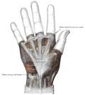
Palm of left hand, showing position of skin creases and bones, and surface markings for the volar arches.
Details
Source Ulnar (primarily), Superficial palmar branch of the radial artery
Branches Common palmar digital
Vein Superficial palmar venous arch
Identifiers
Latin arcus palmaris superficialis,
arcus volaris superficialis
TA98 A12.2.09.056
TA2 4671
FMA 22834
Anatomical terminology

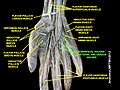
The superficial palmar arch is formed predominantly by the ulnar artery, with a contribution from the superficial palmar branch of the radial artery. However, in some individuals the contribution from the radial artery might be absent, and instead anastomoses with either the princeps pollicis artery, the radialis indicis artery, or the median artery, the former two of which are branches from the radial artery.

Contents

Alternative names for this arterial arch are: superficial volar arch, [1] superficial ulnar arch, arcus palmaris superficialis, [2] or arcus volaris superficialis. [3]

The arch passes across the palm in a curve (Boeckel's line) with its convexity downward,

If one were to fully extend the thumb, the superficial palmar arch would lie approximately 1 cm distal from a line drawn between the first web space to the Hook of Hamate (Kaplan's Cardinal Line). The superficial palmar arch extends more distally than the deep palmar arch. The connection between the deep and superficial palmar arterial arches is an example of anastomosis, and can be tested for using Allen's test.

Three common palmar digital arteries arise from the arch, proceeding down on the second, third, and fourth lumbrical muscles, respectively. They each receive a contribution from a palmar metacarpal artery. Near the level of the metacarpophalangeal joints, each common palmar digital artery divides into two proper palmar digital arteries.

Four digital branches arise from this palmar arch that supplies the medial/ulnar 3 1/2 fingers.

See also

Additional images

Footnotes and references

  1. Palmar and volar may be used synonymously, but volar is less common.
  2. This is the official and international Latin term as defined by the Terminologia Anatomica (TA), but in English speaking countries and especially the US, superficial palmar arch is more commonly used.
  3. Again, palmar and volar may be used synonymously, but arcus volaris superficialis does not occur in the TA, and can therefore be considered deprecated.

Related Research Articles

<span class="mw-page-title-main">Median nerve</span> Nerve of the upper limb

The median nerve is a nerve in humans and other animals in the upper limb. It is one of the five main nerves originating from the brachial plexus.

<span class="mw-page-title-main">Anatomical snuffbox</span> Indent on back of hand between tendons

The anatomical snuff box or snuffbox or foveola radialis is a triangular deepening on the radial, dorsal aspect of the hand—at the level of the carpal bones, specifically, the scaphoid and trapezium bones forming the floor. The name originates from the use of this surface for placing and then sniffing powdered tobacco, or "snuff." It is sometimes referred to by its French name tabatière.

<span class="mw-page-title-main">Ulnar nerve</span> Nerve which runs near the ulna bone

In human anatomy, the ulnar nerve is a nerve that runs near the ulna bone. The ulnar collateral ligament of elbow joint is in relation with the ulnar nerve. The nerve is the largest in the human body unprotected by muscle or bone, so injury is common. This nerve is directly connected to the little finger, and the adjacent half of the ring finger, innervating the palmar aspect of these fingers, including both front and back of the tips, perhaps as far back as the fingernail beds.

Palmar may refer to:

<span class="mw-page-title-main">Upper limb</span> Consists of the arm, forearm, and hand

The upper limbs or upper extremities are the forelimbs of an upright-postured tetrapod vertebrate, extending from the scapulae and clavicles down to and including the digits, including all the musculatures and ligaments involved with the shoulder, elbow, wrist and knuckle joints. In humans, each upper limb is divided into the arm, forearm and hand, and is primarily used for climbing, lifting and manipulating objects.

<span class="mw-page-title-main">Radial artery</span> Large forearm artery

In human anatomy, the radial artery is the main artery of the lateral aspect of the forearm.

<span class="mw-page-title-main">Ulnar artery</span> Artery of the forearm

The ulnar artery is the main blood vessel, with oxygenated blood, of the medial aspects of the forearm. It arises from the brachial artery and terminates in the superficial palmar arch, which joins with the superficial branch of the radial artery. It is palpable on the anterior and medial aspect of the wrist.

<span class="mw-page-title-main">Flexor pollicis brevis muscle</span> Muscle in the thenar compartment

The flexor pollicis brevis is a muscle in the hand that flexes the thumb. It is one of three thenar muscles. It has both a superficial part and a deep part.

<span class="mw-page-title-main">Deep palmar arch</span>

The deep palmar arch is an arterial network found in the palm. It is usually primarily formed from the terminal part of the radial artery. The ulnar artery also contributes through an anastomosis. This is in contrast to the superficial palmar arch, which is formed predominantly by the ulnar artery.

<span class="mw-page-title-main">Palmar metacarpal arteries</span>

The palmar metacarpal arteries are three or four arteries that arise from the convexity of the deep palmar arch.

<span class="mw-page-title-main">Dorsal carpal arch</span>

The dorsal carpal arch is an anatomical term for the combination (anastomosis) of dorsal carpal branch of the radial artery and the dorsal carpal branch of the ulnar artery near the back of the wrist.

<span class="mw-page-title-main">Common palmar digital arteries</span>

Three common palmar digital arteries arise from the convexity of the superficial palmar arch and proceed distally on the second, third, and fourth lumbricales muscles.

In anatomy, arterial tree is used to refer to all arteries and/or the branching pattern of the arteries. This article regards the human arterial tree. Starting from the aorta:

<span class="mw-page-title-main">Palmar carpal arch</span>

The palmar carpal arch is a joining of an artery to an artery, a circulatory anastomosis, known as an arterio-arterial anastomosis. The two connected arteries are the palmar carpal branch of the radial artery and the palmar carpal branch of the ulnar artery.

<span class="mw-page-title-main">Proper palmar digital arteries</span>

The proper palmar digital arteries travel along the sides of the phalanges, each artery lying just below its corresponding digital nerve.

<span class="mw-page-title-main">Dorsal carpal branch of the radial artery</span>

The dorsal carpal branch of the radial artery is a small vessel which arises beneath the extensor tendons of the thumb; crossing the carpus transversely toward the medial border of the hand, it anastomoses with the dorsal carpal branch of the ulnar artery.

<span class="mw-page-title-main">Carpal tunnel</span> Structure of human wrist

In the human body, the carpal tunnel or carpal canal is the passageway on the palmar side of the wrist that connects the forearm to the hand.

<span class="mw-page-title-main">Superficial venous palmar arch</span>

The superficial palmar venous arch consists of a pair of venae comitantes accompanying the superficial palmar arch. It receives the common palmar digital veins. It drains into the superficial ulnar radial and superficial radial veins, and the median antebrachial vein.

<span class="mw-page-title-main">Hand</span> Extremity at the end of an arm or forelimb

A hand is a prehensile, multi-fingered appendage located at the end of the forearm or forelimb of primates such as humans, chimpanzees, monkeys, and lemurs. A few other vertebrates such as the koala are often described as having "hands" instead of paws on their front limbs. The raccoon is usually described as having "hands" though opposable thumbs are lacking.

Volar arteries may refer to:

References

PD-icon.svgThis article incorporates text in the public domain from page 598 of the 20th edition of Gray's Anatomy (1918)