The brachial artery is the major blood vessel of the (upper) arm. It is the continuation of the axillary artery beyond the lower margin of teres major muscle. It continues down the ventral surface of the arm until it reaches the cubital fossa at the elbow. It then divides into the radial and ulnar arteries which run down the forearm. In some individuals, the bifurcation occurs much earlier and the ulnar and radial arteries extend through the upper arm. The pulse of the brachial artery is palpable on the anterior aspect of the elbow, medial to the tendon of the biceps, and, with the use of a stethoscope and sphygmomanometer, often used to measure the blood pressure.
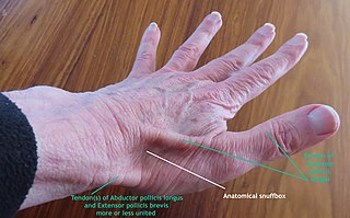
The anatomical snuff box or snuffbox or foveola radialis is a triangular deepening on the radial, dorsal aspect of the hand—at the level of the carpal bones, specifically, the scaphoid and trapezium bones forming the floor. The name originates from the use of this surface for placing and then sniffing powdered tobacco, or "snuff." It is sometimes referred to by its French name tabatière.
In human anatomy, extensor carpi radialis brevis is a muscle in the forearm that acts to extend and abduct the wrist. It is shorter and thicker than its namesake extensor carpi radialis longus which can be found above the proximal end of the extensor carpi radialis brevis.

In human anatomy, the ulnar nerve is a nerve that runs near the ulna bone. The ulnar collateral ligament of elbow joint is in relation with the ulnar nerve. The nerve is the largest in the human body unprotected by muscle or bone, so injury is common. This nerve is directly connected to the little finger, and the adjacent half of the ring finger, innervating the palmar aspect of these fingers, including both front and back of the tips, perhaps as far back as the fingernail beds.

The radius or radial bone is one of the two large bones of the forearm, the other being the ulna. It extends from the lateral side of the elbow to the thumb side of the wrist and runs parallel to the ulna. The ulna is longer than the radius, but the radius is thicker. The radius is a long bone, prism-shaped and slightly curved longitudinally.

The triquetral bone is located in the wrist on the medial side of the proximal row of the carpus between the lunate and pisiform bones. It is on the ulnar side of the hand, but does not directly articulate with the ulna. Instead, it is connected to and articulates with the ulna through the Triangular fibrocartilage disc and ligament, which forms part of the ulnocarpal joint capsule. It connects with the pisiform, hamate, and lunate bones. It is the 2nd most commonly fractured carpal bone.

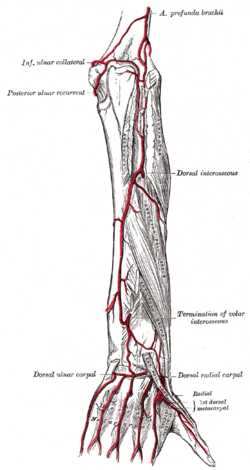
In human anatomy, the radial artery is the main artery of the lateral aspect of the forearm.
The ulnar artery is the main blood vessel, with oxygenated blood, of the medial aspects of the forearm. It arises from the brachial artery and terminates in the superficial palmar arch, which joins with the superficial branch of the radial artery. It is palpable on the anterior and medial aspect of the wrist.
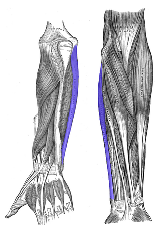
The flexor carpi ulnaris (FCU) is a muscle of the forearm that flexes and adducts at the wrist joint.

In human anatomy, the adductor pollicis muscle is a muscle in the hand that functions to adduct the thumb. It has two heads: transverse and oblique.

In human anatomy, the dorsal interossei of the foot are four muscles situated between the metatarsal bones.

In human anatomy, the extensor pollicis brevis (EPB) is a skeletal muscle on the dorsal side of the forearm. It lies on the medial side of, and is closely connected with, the abductor pollicis longus. The extensor pollicis brevis belongs to the deep group of the posterior fascial compartment of the forearm. It is a part of the lateral border of the anatomical snuffbox.

The flexor retinaculum is a fibrous band on the palmar side of the hand near the wrist. It arches over the carpal bones of the hands, covering them and forming the carpal tunnel.
The deep palmar arch is an arterial network found in the palm. It is usually primarily formed from the terminal part of the radial artery. The ulnar artery also contributes through an anastomosis. This is in contrast to the superficial palmar arch, which is formed predominantly by the ulnar artery.

The superficial branch of the radial nerve passes along the front of the radial side of the forearm to the commencement of its lower third. It is a sensory nerve.
The princeps pollicis artery, or principal artery of the thumb, arises from the radial artery just as it turns medially towards the deep part of the hand; it descends between the first dorsal interosseous muscle and the oblique head of the adductor pollicis, along the medial side of the first metacarpal bone to the base of the proximal phalanx, where it lies beneath the tendon of the flexor pollicis longus muscle and divides into two branches.

The dorsal carpal branch of the radial artery is a small vessel which arises beneath the extensor tendons of the thumb; crossing the carpus transversely toward the medial border of the hand, it anastomoses with the dorsal carpal branch of the ulnar artery.

In the human body, the carpal tunnel or carpal canal is a flattened body cavity on the flexor (palmar/volar) side of the wrist, bounded by the carpal bones and flexor retinaculum. It forms the passageway that transmits the median nerve and the tendons of the extrinsic flexor muscles of the hand from the forearm to the hand. There are described cases of the anatomical variant median artery occurrence.

The extrinsic extensor muscles of the hand are located in the back of the forearm and have long tendons connecting them to bones in the hand, where they exert their action. Extrinsic denotes their location outside the hand. Extensor denotes their action which is to extend, or open flat, joints in the hand. They include the extensor carpi radialis longus (ECRL), extensor carpi radialis brevis (ECRB), extensor digitorum (ED), extensor digiti minimi (EDM), extensor carpi ulnaris (ECU), abductor pollicis longus (APL), extensor pollicis brevis (EPB), extensor pollicis longus (EPL), and extensor indicis (EI).

The muscles of the thumb are nine skeletal muscles located in the hand and forearm. The muscles allow for flexion, extension, adduction, abduction and opposition of the thumb. The muscles acting on the thumb can be divided into two groups: The extrinsic hand muscles, with their muscle bellies located in the forearm, and the intrinsic hand muscles, with their muscles bellies located in the hand proper.