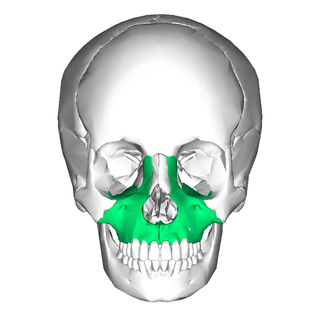
In vertebrates, the maxilla is the upper fixed bone of the jaw formed from the fusion of two maxillary bones. In humans, the upper jaw includes the hard palate in the front of the mouth. The two maxillary bones are fused at the intermaxillary suture, forming the anterior nasal spine. This is similar to the mandible, which is also a fusion of two mandibular bones at the mandibular symphysis. The mandible is the movable part of the jaw.

In the human skull, the zygomatic bone, also called cheekbone or malar bone, is a paired irregular bone, situated at the upper and lateral part of the face and forming part of the lateral wall and floor of the orbit, of the temporal fossa and the infratemporal fossa. It presents a malar and a temporal surface; four processes, and four borders.

The sphenoid bone is an unpaired bone of the neurocranium. It is situated in the middle of the skull towards the front, in front of the basilar part of the occipital bone. The sphenoid bone is one of the seven bones that articulate to form the orbit. Its shape somewhat resembles that of a butterfly or bat with its wings extended.

In anatomy, the palatine bones are two irregular bones of the facial skeleton in many animal species, located above the uvula in the throat. Together with the maxilla, they comprise the hard palate.

The lacrimal bones are two small and fragile bones of the facial skeleton; they are roughly the size of the little fingernail and situated at the front part of the medial wall of the orbit. They each have two surfaces and four borders. Several bony landmarks of the lacrimal bones function in the process of lacrimation. Specifically, the lacrimal bones help form the nasolacrimal canal necessary for tear translocation. A depression on the anterior inferior portion of one bone, the lacrimal fossa, houses the membranous lacrimal sac. Tears, from the lacrimal glands, collect in this sac during excessive lacrimation. The fluid then flows through the nasolacrimal duct and into the nasopharynx. This drainage results in what is commonly referred to a runny nose during excessive crying or tear production. Injury or fracture of the lacrimal bone can result in posttraumatic obstruction of the lacrimal pathways.

In anatomy, the orbit is the cavity or socket/hole of the skull in which the eye and its appendages are situated. "Orbit" can refer to the bony socket, or it can also be used to imply the contents. In the adult human, the volume of the orbit is about 28 millilitres, of which the eye occupies 6.5 ml. The orbital contents comprise the eye, the orbital and retrobulbar fascia, extraocular muscles, cranial nerves II, III, IV, V, and VI, blood vessels, fat, the lacrimal gland with its sac and duct, the eyelids, medial and lateral palpebral ligaments, cheek ligaments, the suspensory ligament, septum, ciliary ganglion and short ciliary nerves.

The middle meningeal artery is typically the third branch of the first portion of the maxillary artery. After branching off the maxillary artery in the infratemporal fossa, it runs through the foramen spinosum to supply the dura mater and the calvaria. The middle meningeal artery is the largest of the three (paired) arteries that supply the meninges, the others being the anterior meningeal artery and the posterior meningeal artery.

The lacrimal glands are paired exocrine glands, one for each eye, found in most terrestrial vertebrates and some marine mammals, that secrete the aqueous layer of the tear film. In humans, they are situated in the upper lateral region of each orbit, in the lacrimal fossa of the orbit formed by the frontal bone. Inflammation of the lacrimal glands is called dacryoadenitis. The lacrimal gland produces tears which are secreted by the lacrimal ducts, and flow over the ocular surface, and then into canals that connect to the lacrimal sac. From that sac, the tears drain through the lacrimal duct into the nose.

The orbital or horizontal part of the frontal bone consists of two thin triangular plates, the orbital plates, which form the vaults of the orbits, and are separated from one another by a median gap, the ethmoidal notch.

In human anatomy, the pterygopalatine fossa is a fossa in the skull. A human skull contains two pterygopalatine fossae—one on the left side, and another on the right side. Each fossa is a cone-shaped paired depression deep to the infratemporal fossa and posterior to the maxilla on each side of the skull, located between the pterygoid process and the maxillary tuberosity close to the apex of the orbit. It is the indented area medial to the pterygomaxillary fissure leading into the sphenopalatine foramen. It communicates with the nasal and oral cavities, infratemporal fossa, orbit, pharynx, and middle cranial fossa through eight foramina.

The greater wing of the sphenoid bone, or alisphenoid, is a bony process of the sphenoid bone, positioned in the skull behind each eye. There is one on each side, extending from the side of the body of the sphenoid and curving upward, laterally, and backward.
The lesser wings of the sphenoid or orbito-sphenoids are two thin triangular plates, which arise from the upper and anterior parts of the body, and, projecting lateralward, end in sharp points [Fig. 1].

The optic foramen is the opening to the optic canal. The canal is located in the sphenoid bone; it is bounded medially by the body of the sphenoid and laterally by the lesser wing of the sphenoid.

In human anatomy, the infraorbital foramen is one of two small holes in the skull's upper jawbone, located below the eye socket and to the left and right of the nose. Both holes are used for blood vessels and nerves. In anatomical terms, it is located below the infraorbital margin of the orbit. It transmits the infraorbital artery and vein, and the infraorbital nerve, a branch of the maxillary nerve. It is typically 6.10 to 10.9 mm from the infraorbital margin.

The middle cranial fossa is formed by the sphenoid bones, and the temporal bones. It lodges the temporal lobes, and the pituitary gland. It is deeper than the anterior cranial fossa, is narrow medially and widens laterally to the sides of the skull. It is separated from the posterior cranial fossa by the clivus and the petrous crest.
The canal containing the nasolacrimal duct is called the nasolacrimal canal.

The pterygoid canal is a passage in the sphenoid bone of the skull leading from just anterior to the foramen lacerum in the middle cranial fossa to the pterygopalatine fossa.

The sphenoidal process of palatine bone is a thin, superomedially directed plate of bone. It is smaller and more inferior compared to the orbital process of palatine bone.

On the nasal surface of the body of the maxilla, in front of the opening of the sinus is a deep groove, the lacrimal groove, which is converted into the nasolacrimal canal, by the lacrimal bone and inferior nasal concha; this canal opens into the inferior meatus of the nose and transmits the nasolacrimal duct.

The following outline is provided as an overview of and topical guide to human anatomy:
