Vulvar cancer

Last updated
Vulvar cancer
The Principles and practice of gynecology - for students and practitioners (1904) (14581300578).jpg
Drawing of vulvar cancer
Specialty Gynecology
Symptoms Lump, itchiness, changes in the skin, or bleeding of the vulva [1]
Usual onsetAfter the age of 45 [2]
Types Squamous cell cancer, adenocarcinoma, melanoma, sarcoma, basal cell carcinoma. [3]
Risk factors Vulvar intraepithelial neoplasia (VIN), HPV infection, genital warts, smoking, many sexual partners [1] [3]
Diagnostic method Physical examination, tissue biopsy [1]
Differential diagnosis Lichen sclerosus, hyperplasia [4]
Prevention HPV vaccination [5]
TreatmentSurgery, radiation therapy, chemotherapy, biologic therapy [1]
Prognosis Five-year survival ~ 71% (US 2015) [2]
Frequency44,200 (2018) [6]
Deaths15,200 (2018) [6]

Vulvar cancer is a cancer of the vulva, the outer portion of the female genitals. [1] It most commonly affects the labia majora. [1] Less often, the labia minora, clitoris, or Bartholin's glands are affected. [1] Symptoms include a lump, itchiness, changes in the skin, or bleeding from the vulva. [1]

Contents

Risk factors include vulvar intraepithelial neoplasia (VIN), HPV infection, genital warts, smoking, and many sexual partners. [1] [3] Most vulvar cancers are squamous cell cancers. [4] Other types include adenocarcinoma, melanoma, sarcoma, and basal cell carcinoma. [3] Diagnosis is suspected based on physical examination and confirmed by tissue biopsy. [1] Routine screening is not recommended. [3]

Prevention may include HPV vaccination. [5] Standard treatments may include surgery, radiation therapy, chemotherapy, and biologic therapy. [1] Vulvar cancer newly affected about 44,200 people and resulted in 15,200 deaths globally in 2018. [6] In the United States, it newly occurred in about 6,070 people with 1,280 deaths a year. [2] Onset is typically after the age of 45. [2] The five-year survival rate for vulvar cancer is around 71% as of 2015. [2] Outcomes, however, are affected by whether spread has occurred to lymph nodes. [4]

Signs and symptoms

Drawing of cancer of the clitoris with spread to the groin Gynecology - (1918) (14750388526).jpg
Drawing of cancer of the clitoris with spread to the groin

The signs and symptoms can include:

Typically, a lesion presents in the form of a lump or ulcer on the labia majora and may be associated with itching, irritation, local bleeding or discharge, in addition to pain with urination or pain during sexual intercourse. [8] The labia minora, clitoris, perineum and mons pubis are less commonly involved. [9] Due to modesty or embarrassment, people may put off seeing a doctor. [10]

Melanomas tend to display the typical asymmetry, uneven borders and dark discoloration as do melanomas in other parts of the body.

Adenocarcinoma can arise from the Bartholin's gland and present with a painful lump. [11]

Causes

Two main pathophysiological pathways are currently understood to contribute to development of vulvar cancer—human papillomavirus (HPV) infection and chronic inflammation or autoimmunity affecting the vulvar area. [12] [13] [14]

HPV DNA can be found in up to 87% of vulvar intraepithelial neoplasia (VIN) and 29% of invasive vulvar cancers; HPV 16 is the most commonly detected subtype in VIN and vulvar cancer, followed by HPV 33 and HPV 18. [15] VIN is a superficial lesion of the skin that has not invaded the basement membrane—or a pre-cancer. [16] VIN may progress to carcinoma in situ and, eventually, squamous cell cancer.

Chronic inflammatory conditions of the vulva that may be precursors to vulvar cancer include lichen sclerosus, which can predispose to differentiated VIN. [17] [18]

Risk factors

Risk factors for vulvar cancer are largely related to the causal pathways above, involving exposure or infection with the HPV virus and/or acquired or innate auto-immunity. [19] [20]

Diagnosis

Examination of the vulva is part of the gynecologic evaluation and should include a thorough inspection of the perineum, including areas around the clitoris and urethra, and palpation of the Bartholin's glands. [21] The exam may reveal an ulceration, lump or mass in the vulvar region. Any suspicious lesions need to be sampled, or biopsied. This can generally be done in an office setting under local anesthesia. Small lesions can be removed under local anesthesia as well. Additional evaluation may include a chest X-ray, an intravenous pyelogram, cystoscopy or proctoscopy, as well as blood counts and metabolic assessment.

Types

Depending on the cellular origin, different histologic cancer subtypes may arise in vulvar structures. [22] [23]

Squamous cell carcinoma

A recent analysis of the Surveillance, Epidemiology and End Results (SEER) registry of the US National Cancer Institute has shown that squamous cell carcinoma accounts for approximately 75% of all vulvar cancers. [22] These lesions originate from epidermal squamous cells, the most common type of skin cell. Carcinoma-in-situ is a precursor lesion of squamous cell cancer that does not invade through the basement membrane. There are two types of precursor lesions:

  • Usual-type vulvar intraepithelial neoplasia (uVIN), which is associated with human papillomavirus (HPV) and often affects younger women. This precursor lesion progresses to basaloid or warty squamous cell carcinoma in approximately 6%. [22]
  • Differentiated vulvar intraepithelial neoplasia (dVIN), which is associated with chronic skin conditions including lichen sclerosus and lichen planus and typically affects older women. This lesion progresses to keratinizing squamous cell carcinoma in approximately 33%. [22]

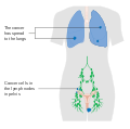
Squamous lesions tend to arise in a single site and occur most commonly in the vestibule. [24] They grow by local extension and spread via the local lymph system. The lymphatics of the labia drain to the upper vulva and mons pubis, then to both superficial and deep inguinal and femoral lymph nodes. The last deep femoral node is called the Cloquet's node. [24] Spread beyond this node reaches the lymph nodes of the pelvis. The tumor may also invade nearby organs such as the vagina, urethra, and rectum and spread via their lymphatics.

A verrucous carcinoma of the vulva is a rare subtype of squamous cell cancer and tends to appear as a slowly growing wart. Verrucous vulvar cancers tend to have a good overall prognosis, as these lesions hardly ever spread to regional lymph nodes or metastasize. [22] [25]

Basal cell carcinoma

Basal cell carcinoma account for approximately 8% of all vulvar cancers. It typically affects women in the 7th and 8th decade of life. [22] These tend to be slow-growing lesions on the labia majora but can occur anywhere on the vulva. Their behavior is similar to basal cell cancers in other locations. They often grow locally and have low risk for deep invasion or metastasis.

Treatment involves local excision, but these lesions have a tendency to recur if not completely removed.

Melanoma

Melanoma is the third most common type and accounts for 6% of all vulvar cancers. [22] These lesions arise from melanocytes, the cells that give skin color. The median age at diagnosis is 68 years; however, an analysis of the Surveillance, Epidemiology and End Results (SEER) registry of the US National Cancer Institute has shown that it has been diagnosed in girls as young as 10 years and women up to 107 years. [22] [26]

The underlying biology of vulvar melanoma differs significantly from skin melanomas and mutational analyses have shown only 8% harbor a BRAF mutation compared to 70% of skin melanomas. [27] KIT mutations, however are significantly more common in vulvar melanoma. [22] [27] This has a direct impact on the medical treatment of vulvar melanomas: BRAF-inhibitors that are commonly used in the treatment of skin melanomas, play a minor role in vulvar melanomas. However, vulvar melanomas frequently express PD-L1 and checkpoint inhibitors (including CTLA-4 inhibitors and PD-1 inhibitors) are effective in the treatment of advanced-stage vulvar melanoma. [28] In recurrent melanoma, tyrosine kinase inhibitors may be used in those patients with a KIT mutation. [22] [27]

Based on histology, there are different subtypes of vulvar melanoma: superficial spreading, nodular, acral lentigous and amelanotic melanoma. Vulvar melanomas are unique in that they are staged using the AJCC cancer staging for melanoma instead of the FIGO staging system. [29]

Diagnosis of vulvar melanoma is often delayed and approximately 32% of women already have regional lymph node involvement or distant metastases at the time of diagnosis. [24] [29] Lymph node metastases and high mitotic count are indicators of poor outcome. [29] The overall prognosis is poor and significantly worse than in skin melanomas: The median overall survival is 53 months. [29] [28]

Bartholin gland carcinoma

The Bartholin gland is a rare malignancy and usually occurs in women in their mid-sixties.

Other lesions

Other forms of vulvar cancer include invasive Extramammary Paget's disease, adenocarcinoma (of the Bartholin glands, for example) and sarcoma. [22] [30]

Staging

Anatomical staging supplemented preclinical staging starting in 1988. FIGO's revised TNM classification system uses tumor size (T), lymph node involvement (N) and presence or absence of metastasis (M) as criteria for staging. Stages I and II describe the early stages of vulvar cancer that still appear to be confined to the site of origin. Stage III cancers include greater disease extension to neighboring tissues and inguinal lymph nodes on one side. Stage IV indicates metastatic disease to inguinal nodes on both sides or distant metastases. [31]

Differential diagnosis

Other cancerous lesions in the differential diagnosis include Paget's disease of the vulva and vulvar intraepithelial neoplasia (VIN). Non-cancerous vulvar diseases include lichen sclerosus, squamous cell hyperplasia, and vulvar vestibulitis. A number of diseases cause infectious lesions including herpes genitalis, human papillomavirus, syphilis, chancroid, granuloma inguinale, and lymphogranuloma venereum.

Treatment

Diagram of the incisions made in a vulvectomy, a treatment for vulvar cancer Diagram of a 3 in 1 incision vulvectomy CRUK 018.svg
Diagram of the incisions made in a vulvectomy, a treatment for vulvar cancer

Surgery is a mainstay of therapy depending on anatomical staging and is usually reserved for cancers that have not spread beyond the vulva. [31] Surgery may involve a wide local excision (excision of the tumor with a safety-margin of healthy tissue, that ensures complete removal of the tumor), radical partial vulvectomy, or radical complete vulvectomy with removal of vulvar tissue, inguinal and femoral lymph nodes. [22] [26] In cases of early vulvar cancer, the surgery may be less extensive and consist of wide excision or a simple vulvectomy. Surgery is significantly more extensive when the cancer has spread to nearby organs such as the urethra, vagina, or rectum. Complications of surgery include wound infection, sexual dysfunction, edema and thrombosis, as well as lymphedema secondary to dissected lymph nodes. [32]

Sentinel lymph node (SLN) dissection is the identification of the main lymph node(s) draining the tumor, with the aim of removing as few nodes as possible, decreasing the risk of adverse effects. Location of the sentinel node(s) may require the use of technetium(99m)-labeled nano-colloid, or a combination of technetium and 1% isosulfan blue dye, wherein the combination may reduce the number of women with "'missed"' groin node metastases compared with technetium only. [32]

Radiation therapy may be used in more advanced vulvar cancer cases when disease has spread to the lymph nodes and/or pelvis. It may be performed before or after surgery. In early vulvar cancer, primary radiotherapy to the groin results in less morbidity but may be linked with a higher risk of groin recurrence and reduced survival compared to surgery. [33] Chemotherapy is not usually used as primary treatment but may be used in advanced cases with spread to the bones, liver or lungs. It may also be given at a lower dose together with radiation therapy. [34] Checkpoint inhibitors may be given in melanoma of the vulva. [28]

There is no significant difference in overall survival or treatment‐related adverse effects in women with locally advanced vulval cancer when comparing primary chemoradiation or neoadjuvant chemoradiation with primary surgery. There is a need for good quality studies comparing various primary treatments. [35]

Women with vulvar cancer should have routine follow-up and exams with their oncologist, often every three months for the first 2–3 years after treatment. They should not have routine surveillance imaging to monitor the cancer unless new symptoms appear or tumor markers begin rising. [36] Imaging without these indications is discouraged because it is unlikely to detect a recurrence or improve survival and is associated with its own side effects and financial costs. [36]

Prognosis

Overall, five-year survival rates for vulvar cancer are around 78% [24] but may be affected by individual factors including cancer stage, cancer type, patient age and general medical health. Five-year survival is greater than 90% for patients with stage I lesions but decreases to 20% when pelvic lymph nodes are involved. Lymph node involvement is the most important predictor of prognosis. [37]

Prognosis depends on the stage of cancer, which refers the amount and spread of cancer in the body. [38] The stages are broken into four categories. Stage one also called "localized" and is when the cancer is limited to one part of the body. [38] This has the highest survival rate of 59%. [38] When the cancer starts to spread this is referred to "distant" or "regional", this stage usually involves the cancer being spread to the lymph nodes. [38] This survival rate is 29%. The third stage is when the cancer has metastasized and spread throughout the body, this is the lowest survival rate of 6%. When vulvar cancer is caught early that is when the survival rate is at its highest. [38]

Epidemiology

Vulvar cancer newly affected about 44,200 people and resulted in 15,200 deaths globally in 2018. [6]

Vulvar cancer can be split up into two types. One starts as an infection by human papillomavirus, which leads to vulvar intraepithelial neoplasia (VIN) and potentially on to vulvar cancer. [39] This is most common in younger women, predominantly under the age of 40. [39] The second type is vulvar non-neoplastic epithelial disorders (VNED). This is most common in older women, due to the increased risk for developing cellular atypia which in turn leads to cancer. [39]

United Kingdom

Vulvar cancer causes less than 1% of all cancer cases and deaths but around 6% of all gynecologic cancers diagnosed in the UK. Around 1,200 women were diagnosed with the disease in 2011, and 400 women died in 2012. [40] In the United Kingdom 7 out of 10 vulval cancer patients have major surgical resection as part of their cancer treatment. [41] 22% of patients use radiotherapy and only 7% use chemotherapy as a treatment plan. [41] There are very high survival rates, patients diagnosed with vulvar cancer have an 82% of living more than one year, a 64% chance of living at least five years and a 53% chance of living ten or more years. [41] The rate of survival increases dependent on age of patient and the stage the cancer was found in.

United States

In the United States, it newly occurred in about 6,070 people with 1,280 deaths a year. [2] It makes up about 0.3% of new cancer cases, [2] and 5% of gynecologic cancers in the United States. [42] Vulvar cancer cases have been rising in the United States at an increase of 0.6% each year for the past ten years. [38]

See also

Related Research Articles

<span class="mw-page-title-main">Cervical cancer</span> Cancer arising from the cervix

Cervical cancer is a cancer arising from the cervix or in the any layer of the wall of the cervix. It is due to the abnormal growth of cells that have the ability to invade or spread to other parts of the body. Early on, typically no symptoms are seen. Later symptoms may include abnormal vaginal bleeding, pelvic pain or pain during sexual intercourse. While bleeding after sex may not be serious, it may also indicate the presence of cervical cancer.

<span class="mw-page-title-main">Bartholin's gland</span> Mucous glands located near the vaginal opening

The Bartholin's glands are two pea-sized compound alveolar glands located slightly posterior and to the left and right of the opening of the vagina. They secrete mucus to lubricate the vagina.

Penile cancer, or penile carcinoma, is a cancer that develops in the skin or tissues of the penis. Symptoms may include abnormal growth, an ulcer or sore on the skin of the penis, and bleeding or foul smelling discharge.

<span class="mw-page-title-main">Anal cancer</span> Medical condition

Anal cancer is a cancer which arises from the anus, the distal opening of the gastrointestinal tract. Symptoms may include bleeding from the anus or a lump near the anus. Other symptoms may include pain, itchiness, or discharge from the anus. A change in bowel movements may also occur.

<span class="mw-page-title-main">Cervical intraepithelial neoplasia</span> Medical condition

Cervical intraepithelial neoplasia (CIN), also known as cervical dysplasia, is the abnormal growth of cells on the surface of the cervix that could potentially lead to cervical cancer. More specifically, CIN refers to the potentially precancerous transformation of cells of the cervix.

<span class="mw-page-title-main">Vulvar intraepithelial neoplasia</span> Medical condition

Vulvar intraepithelial neoplasia (VIN) refers to particular changes that can occur in the skin that covers the vulva. VIN is an intraepithelial neoplasia, and can disappear without treatment. VINs are benign but if the changes become more severe, there is a chance of cancer developing after many years, and so it is referred to as a precancerous condition.

Vaginal cancer is an extraordinarily rare form of cancer that develops in the tissue of the vagina. Primary vaginal cancer originates from the vaginal tissue – most frequently squamous cell carcinoma, but primary vaginal adenocarcinoma, sarcoma, and melanoma have also been reported – while secondary vaginal cancer involves the metastasis of a cancer that originated in a different part of the body. Secondary vaginal cancer is more common. Signs of vaginal cancer may include abnormal vaginal bleeding, dysuria, tenesmus, or pelvic pain, though as many as 20% of women diagnosed with vaginal cancer are asymptomatic at the time of diagnosis. Vaginal cancer occurs more frequently in women over age 50, and the mean age of diagnosis of vaginal cancer is 60 years. It often can be cured if found and treated in early stages. Surgery alone or surgery combined with pelvic radiation is typically used to treat vaginal cancer.

Gynecologic oncology is a specialized field of medicine that focuses on cancers of the female reproductive system, including ovarian cancer, uterine cancer, vaginal cancer, cervical cancer, and vulvar cancer. As specialists, they have extensive training in the diagnosis and treatment of these cancers.

Gynecologic cancer is a type of cancer that affects the female reproductive system, including ovarian cancer, uterine cancer, vaginal cancer, cervical cancer, and vulvar cancer.

<span class="mw-page-title-main">Oropharyngeal cancer</span> Pharynx cancer that is located in the oropharynx

Oropharyngeal cancer, also known as oropharyngeal squamous cell carcinoma and tonsil cancer, is a disease in which abnormal cells with the potential to both grow locally and spread to other parts of the body are found in the oral cavity, in the tissue of the part of the throat (oropharynx) that includes the base of the tongue, the tonsils, the soft palate, and the walls of the pharynx.

Neuroendocrine carcinoma of the cervix is best defined separately:Neuroendocrine: Of, relating to, or involving the interaction between the nervous system and the hormones of the endocrine glands.Carcinoma: An invasive malignant tumor derived from epithelial tissue that tends to metastasize to other areas of the body.

<span class="mw-page-title-main">Vulva</span> External genitalia of the female mammal

In mammals, the vulva consists of the external female genitalia. The human vulva includes the mons pubis, labia majora, labia minora, clitoris, vulval vestibule, urinary meatus, the vaginal opening, hymen, and Bartholin's and Skene's vestibular glands. The urinary meatus is also included as it opens into the vulval vestibule. The vulva includes the entrance to the vagina, which leads to the uterus, and provides a double layer of protection for this by the folds of the outer and inner labia. Pelvic floor muscles support the structures of the vulva. Other muscles of the urogenital triangle also give support.

<span class="mw-page-title-main">Cervical cancer staging</span> Medical condition

Cervical cancer staging is the assessment of cervical cancer to determine the extent of the spread of cancer beyond the cervix. This is important for determining how serious the cancer is and to create the best treatment plan.

Conjunctival squamous cell carcinoma and corneal intraepithelial neoplasia comprise ocular surface squamous neoplasia (OSSN). SCC is the most common malignancy of the conjunctiva in the US, with a yearly incidence of 1–2.8 per 100,000. Risk factors for the disease are exposure to sun, exposure to UVB, and light-colored skin. Other risk factors include radiation, smoking, HPV, arsenic, and exposure to polycyclic hydrocarbons.

A vulvar disease is a particular abnormal, pathological condition that affects part or all of the vulva. Several pathologies are defined. Some can be prevented by vulvovaginal health maintenance.

Lymphatogenous metastasis is the spread of cancer from the vulva, clitoris and Bartholin glands. As these other more superficial cancers grow deeper into the surrounding tissue, the risk of lymphatogenous metastasis increases. Vulvar cancers spread primarily to the inguinofemoral lymph nodes. From these lymph nodes, cancer can spread to the pelvic lymph nodes.

<span class="mw-page-title-main">Bartholin gland carcinoma</span> Medical condition

Bartholin gland carcinoma is a type of cancer of the vulva arising in the Bartholin gland. It typically presents with a painless mass at one side of the vaginal opening in a female of middle-age and older, and can appear similar to a Bartholin cyst. The mass may be big or small, may be deep under skin or appear nearer the surface with overlying ulceration. Average age at presentation is 53-years.

Carcinoma of the tonsil is a type of squamous cell carcinoma. The tonsil is the most common site of squamous cell carcinoma in the oropharynx. It comprises 23.1% of all malignancies of the oropharynx. The tumors frequently present at advanced stages, and around 70% of patients present with metastasis to the cervical lymph nodes. . The most reported complaints include sore throat, otalgia or dysphagia. Some patients may complain of feeling the presence of a lump in the throat. Approximately 20% patients present with a node in the neck as the only symptom.

Vaginal melanoma is a rare malignancy that originates from melanocytes in the vaginal epithelium. It is also known as a melanocytic tumor or as a malignant melanoma. It is aggressive and infrequently cured. The median overall survival is 16 months. Vaginal melanoma accounts 5.5% of all vaginal cancers and only 1% of all melanomas diagnosed in women. Vaginal melanomas are frequently diagnosed in advanced stages of the disease. The prognosis is poor and the most important risk factor is the presence of lymph node metastases.

Vulvar tumors are those neoplasms of the vulva. Vulvar and vaginal neoplasms make up a small percentage (3%) of female genital cancers. They can be benign or malignant. Vulvar neoplasms are divided into cystic or solid lesions and other mixed types. Vulvar cancers are those malignant neoplasms that originate from vulvar epithelium, while vulvar sarcomas develop from non-epithelial cells such as bone, cartilage, fat, muscle, blood vessels, or other connective or supportive tissue. Epithelial and mesenchymal tissue are the origin of vulvar tumors.

References

  1. 1 2 3 4 5 6 7 8 9 10 11 "Vulvar Cancer Treatment". National Cancer Institute. 9 April 2019. Retrieved 31 May 2019.
  2. 1 2 3 4 5 6 7 "Cancer of the Vulva—Cancer Stat Facts". SEER. Retrieved 30 May 2019.
  3. 1 2 3 4 5 Sam A, George J, Mathew B (April 2019). "Less Common Gynecologic Malignancies: An Integrative Review". Seminars in Oncology Nursing. 35 (2): 175–181. doi:10.1016/j.soncn.2019.02.004. PMID   30867101. S2CID   76665631.
  4. 1 2 3 "Vulvar Cancer Treatment". National Cancer Institute. 1 February 2019. Retrieved 31 May 2019.
  5. 1 2 Signorelli C, Odone A, Ciorba V, Cella P, Audisio RA, Lombardi A, et al. (July 2017). "Human papillomavirus 9-valent vaccine for cancer prevention: a systematic review of the available evidence". Epidemiology and Infection. 145 (10): 1962–1982. doi:10.1017/S0950268817000747. PMC   5974698 . PMID   28446260.
  6. 1 2 3 4 "Cancer today". IARC. Retrieved 30 May 2019.
  7. "What Are the Symptoms of Vaginal and Vulvar Cancers?". CDC. 13 March 2014. Retrieved 2016-03-26.
  8. "Vulvar Cancer Treatment". National Cancer Institute. 1980-01-01. Retrieved 2015-04-14.
  9. "What is vulvar cancer?". cancer.org. Retrieved 2015-04-14.
  10. Zacur H, Genadry R, Woodruff JD (April 1980). "The patient-at-risk for development of vulvar cancer". Gynecologic Oncology. 9 (2): 199–208. doi:10.1016/0090-8258(80)90028-1. PMID   7372192.
  11. Copeland LJ, Sneige N, Gershenson DM, McGuffee VB, Abdul-Karim F, Rutledge FN (June 1986). "Bartholin gland carcinoma". Obstetrics and Gynecology. 67 (6): 794–801. doi:10.1097/00006250-198606000-00009. PMID   3010205.
  12. de Koning MN, Quint WG, Pirog EC (March 2008). "Prevalence of mucosal and cutaneous human papillomaviruses in different histologic subtypes of vulvar carcinoma". Modern Pathology. 21 (3): 334–44. doi: 10.1038/modpathol.3801009 . PMID   18192968.
  13. Weberpals JI, Lo B, Duciaume MM, Spaans JN, Clancy AA, Dimitroulakos J, et al. (August 2017). "Vulvar Squamous Cell Carcinoma (VSCC) as Two Diseases: HPV Status Identifies Distinct Mutational Profiles Including Oncogenic Fibroblast Growth Factor Receptor 3". Clinical Cancer Research. 23 (15): 4501–4510. doi: 10.1158/1078-0432.CCR-16-3230 . PMID   28377483.
  14. Halec G, Alemany L, Quiros B, Clavero O, Höfler D, Alejo M, et al. (April 2017). "Biological relevance of human papillomaviruses in vulvar cancer". Modern Pathology. 30 (4): 549–562. doi: 10.1038/modpathol.2016.197 . PMID   28059099.
  15. de Sanjosé S, Alemany L, Ordi J, Tous S, Alejo M, Bigby SM, et al. (November 2013). "Worldwide human papillomavirus genotype attribution in over 2000 cases of intraepithelial and invasive lesions of the vulva". European Journal of Cancer. 49 (16): 3450–61. doi:10.1016/j.ejca.2013.06.033. PMID   23886586.
  16. "What is Vulvar Cancer?". Society of Gynecologic Oncology. 21 November 2012. Retrieved 19 November 2014.
  17. van de Nieuwenhof HP, Bulten J, Hollema H, Dommerholt RG, Massuger LF, van der Zee AG, et al. (February 2011). "Differentiated vulvar intraepithelial neoplasia is often found in lesions, previously diagnosed as lichen sclerosus, which have progressed to vulvar squamous cell carcinoma". Modern Pathology. 24 (2): 297–305. doi: 10.1038/modpathol.2010.192 . PMID   21057461.
  18. Bigby SM, Eva LJ, Fong KL, Jones RW (November 2016). "The Natural History of Vulvar Intraepithelial Neoplasia, Differentiated Type: Evidence for Progression and Diagnostic Challenges". International Journal of Gynecological Pathology. 35 (6): 574–584. doi:10.1097/PGP.0000000000000280. PMID   26974999. S2CID   42163280.
  19. Madsen BS, Jensen HL, van den Brule AJ, Wohlfahrt J, Frisch M (June 2008). "Risk factors for invasive squamous cell carcinoma of the vulva and vagina—population-based case-control study in Denmark". International Journal of Cancer. 122 (12): 2827–34. doi: 10.1002/ijc.23446 . PMID   18348142. S2CID   11542729.
  20. Brinton LA, Thistle JE, Liao LM, Trabert B (May 2017). "Epidemiology of vulvar neoplasia in the NIH-AARP Study". Gynecologic Oncology. 145 (2): 298–304. doi:10.1016/j.ygyno.2017.02.030. PMC   5629039 . PMID   28236455.
  21. "Vulvar Cancer". Gynecologic Neoplasms. Armenian Health Network, Health.am. 2005. Retrieved 2007-11-08.
  22. 1 2 3 4 5 6 7 8 9 10 11 12 Wohlmuth, Christoph; Wohlmuth-Wieser, Iris (December 2019). "Vulvar malignancies: an interdisciplinary perspective". Journal of the German Society of Dermatology. 17 (12): 1257–1276. doi:10.1111/ddg.13995. ISSN   1610-0387. PMC   6972795 . PMID   31829526.
  23. Hoffman, Barbara (2012). Williams Gynecology (2nd. ed.). New York: McGraw-Hill Medical. pp. 794–806. ISBN   9780071716727.
  24. 1 2 3 4 Hoffman B, Schorge J, Schaffer J, Halvorson L, Bradshaw K, Cunningham G (2012). Williams Gynecology (2nd ed.). The McGraw-Hill Company, Inc. ISBN   978-0-07-171672-7.
  25. "American Cancer Society: What is Vulvar Cancer?". www.cancer.org. Retrieved 2014-06-11.
  26. 1 2 Wohlmuth, Christoph; Wohlmuth-Wieser, Iris; May, Taymaa; Vicus, Danielle; Gien, Lilian T.; Laframboise, Stéphane (April 2020). "Malignant Melanoma of the Vulva and Vagina: A US Population-Based Study of 1863 Patients". American Journal of Clinical Dermatology. 21 (2): 285–295. doi:10.1007/s40257-019-00487-x. ISSN   1179-1888. PMC   7125071 . PMID   31784896.
  27. 1 2 3 Wohlmuth, Christoph; Wohlmuth-Wieser, Iris (2021-06-14). "Vulvar Melanoma: Molecular Characteristics, Diagnosis, Surgical Management, and Medical Treatment". American Journal of Clinical Dermatology. 22 (5): 639–651. doi: 10.1007/s40257-021-00614-7 . ISSN   1179-1888. PMC   8421300 . PMID   34125416.
  28. 1 2 3 Wohlmuth, Christoph; Wohlmuth-Wieser, Iris; Laframboise, Stéphane (2020-11-24). "Clinical Characteristics and Treatment Response With Checkpoint Inhibitors in Malignant Melanoma of the Vulva and Vagina". Journal of Lower Genital Tract Disease. 25 (2): 146–151. doi:10.1097/LGT.0000000000000583. ISSN   1526-0976. PMC   7984764 . PMID   33252450.
  29. 1 2 3 4 Wohlmuth, Christoph; Wohlmuth-Wieser, Iris; May, Taymaa; Vicus, Danielle; Gien, Lilian T.; Laframboise, Stéphane (April 2020). "Malignant Melanoma of the Vulva and Vagina: A US Population-Based Study of 1863 Patients". American Journal of Clinical Dermatology. 21 (2): 285–295. doi:10.1007/s40257-019-00487-x. ISSN   1179-1888. PMC   7125071 . PMID   31784896.
  30. Visco AG, Del Priore G (February 1996). "Postmenopausal bartholin gland enlargement: a hospital-based cancer risk assessment". Obstetrics and Gynecology. 87 (2): 286–90. doi:10.1016/0029-7844(95)00404-1. PMID   8559540. S2CID   11159562.
  31. 1 2 International Federation of Gynecologists and Obstetricians (FIGO) (2000). "Staging classification and clinical practice guidelines of gynaecologic cancers" (PDF). Archived from the original (PDF) on 2006-04-23. Retrieved 2006-10-13.
  32. 1 2 Lawrie TA, Patel A, Martin-Hirsch PP, Bryant A, Ratnavelu ND, Naik R, Ralte A (June 2014). "Sentinel node assessment for diagnosis of groin lymph node involvement in vulval cancer". The Cochrane Database of Systematic Reviews. 2014 (6): CD010409. doi:10.1002/14651858.CD010409.pub2. PMC   6457826 . PMID   24970683.
  33. van der Velden J, Fons G, Lawrie TA (May 2011). "Primary groin irradiation versus primary groin surgery for early vulvar cancer". The Cochrane Database of Systematic Reviews. 2011 (5): CD002224. doi:10.1002/14651858.cd002224.pub2. PMC   7154218 . PMID   21563133.
  34. "What are the treatment options?". Society of Gynecologic Oncology. 21 November 2012. Retrieved 19 November 2014.
  35. Shylasree TS, Bryant A, Howells RE (April 2011). "Chemoradiation for advanced primary vulval cancer". The Cochrane Database of Systematic Reviews. 2016 (4): CD003752. doi:10.1002/14651858.cd003752.pub3. PMC   4164938 . PMID   21491387.
  36. 1 2 Society of Gynecologic Oncology (February 2014), "Five Things Physicians and Patients Should Question", Choosing Wisely: an initiative of the ABIM Foundation , Society of Gynecologic Oncology, retrieved 19 February 2013
  37. Farias-Eisner R, Cirisano FD, Grouse D, Leuchter RS, Karlan BY, Lagasse LD, Berek JS (April 1994). "Conservative and individualized surgery for early squamous carcinoma of the vulva: the treatment of choice for stage I and II (T1-2N0-1M0) disease". Gynecologic Oncology. 53 (1): 55–8. doi:10.1006/gyno.1994.1087. PMID   8175023.
  38. 1 2 3 4 5 6 "Cancer of the Vulva—Cancer Stat Facts". SEER. Retrieved 2019-12-03.
  39. 1 2 3 Alkatout I, Schubert M, Garbrecht N, Weigel MT, Jonat W, Mundhenke C, Günther V (March 20, 2015). "Vulvar cancer: epidemiology, clinical presentation, and management options". International Journal of Women's Health. 7: 305–13. doi: 10.2147/IJWH.S68979 . PMC   4374790 . PMID   25848321.
  40. "Vulval cancer statistics". Cancer Research UK. Retrieved 28 October 2014.
  41. 1 2 3 "Vulval cancer statistics". Cancer Research UK. 2015-05-14. Retrieved 2019-12-03.
  42. "Vulvar Cancer Treatment". National Cancer Institute. 1980-01-01. Retrieved 12 May 2015.