Bethesda system

Last updated

The Bethesda system (TBS), officially called The Bethesda System for Reporting Cervical Cytology, is a system for reporting cervical or vaginal cytologic diagnoses, [1] used for reporting Pap smear results. It was introduced in 1988 [2] and revised in 1991, [3] 2001, [1] [4] [5] and 2014. [6] The name comes from the location (Bethesda, Maryland) of the conference, sponsored by the National Institutes of Health, that established the system.

Contents

Since 2010, there is also a Bethesda system used for cytopathology of thyroid nodules, which is called The Bethesda System for Reporting Thyroid Cytopathology (TBSRTC or BSRTC). Like TBS, it was the result of a conference sponsored by the NIH and is published in book editions (currently by Springer). Mentions of "the Bethesda system" without further specification usually refer to the cervical system, unless the thyroid context of a discussion is implicit.

Cervix

Abnormal results include:[ citation needed ]

The results are calculated differently following a Pap smear of the cervix.[ citation needed ]

Squamous cell abnormalities

LSIL: low-grade squamous intraepithelial lesion

Cytopathology of low-grade squamous intraepithelial lesion (LSIL), with main features, as compared to an unremarkable intermediate squamous cell. Pap stain. Cytopathology of low-grade squamous intraepithelial lesion (LSIL).png
Cytopathology of low-grade squamous intraepithelial lesion (LSIL), with main features, as compared to an unremarkable intermediate squamous cell. Pap stain.

A low-grade squamous intraepithelial lesion (LSIL or LGSIL) indicates possible cervical dysplasia. LSIL usually indicates mild dysplasia (CIN 1), more than likely caused by a human papillomavirus infection. It is usually diagnosed following a Pap smear.[ citation needed ]

CIN 1 is the most common and most benign form of cervical intraepithelial neoplasia and usually resolves spontaneously within two years. Because of this, LSIL results can be managed with a simple "watch and wait" philosophy. However, because there is a 12–16% chance of progression to more severe dysplasia, the physician may want to follow the results more aggressively by performing a colposcopy with biopsy. [7] If the dysplasia progresses, treatment may be necessary. Treatment involves removal of the affected tissue, which can be accomplished by LEEP, cryosurgery, cone biopsy, or laser ablation.[ citation needed ]

HSIL: high-grade squamous intraepithelial lesion

HSIL. Pap stain. High-grade squamous intraepithelial lesion.jpg
HSIL. Pap stain.

High-grade squamous intraepithelial lesion (HSIL or HGSIL) indicates moderate or severe cervical intraepithelial neoplasia or carcinoma in situ. It is usually diagnosed following a Pap test. In some cases these lesions can lead to invasive cervical cancer, if not followed appropriately.[ citation needed ]

HSIL does not mean that cancer is present. Of all women with HSIL results, 2% [8] or less [9] have invasive cervical cancer at that time, however about 20% would progress to having invasive cervical cancer without treatment. [10] To combat this progression, HSIL is usually followed by an immediate colposcopy with biopsy to sample or remove the dysplastic tissue. This tissue is sent for pathology testing to assign a histologic classification that is more definitive than a Pap smear result (which is a cytologic finding). HSIL generally corresponds to the histological classification of CIN 2 or 3.[ citation needed ]

HSIL treatment involves the removal or destruction of the affected cells, usually by LEEP. Other methods include cryotherapy, cautery, or laser ablation, but none are performed on pregnant women for fear of disrupting the pregnancy. [11] Any of these procedures is 85% likely to cure the problem.

Glandular cell abnormalities

Adenocarcinoma

Adenocarcinoma. Pap stain. Adenocarcinoma on pap test 1.jpg
Adenocarcinoma. Pap stain.

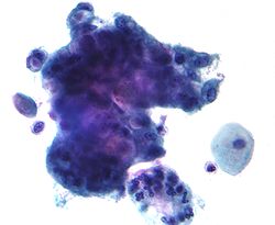
Adenocarcinoma can arise from the endocervix, endometrium and extrauterine sites.[ citation needed ]

AGC

AGC, formerly AGUS, is a term for atypical glandular cells of undetermined significance. [12] Renamed AGC to avoid confusion with ASCUS. [1]

The management of AGC is colposcopy with or without an endometrial biopsy.[ citation needed ]

Thyroid nodules

The Bethesda System for Reporting Thyroid Cytopathology is the system used to report whether the thyroid cytological specimen is benign or malignant on fine-needle aspiration cytology (FNAC). It can be divided into six categories:

Bethesda system
CategoryDescriptionRisk of malignancy [13] Recommendation [13]
INon diagnostic/unsatisfactory-Repeating FNAC with ultrasound-guidance in more than 3 months
IIBenign (colloid and follicular cells)0 - 3%Clinical follow-up
IIIAtypia of undetermined significance/follicular lesion of undetermined significance (follicular or lymphoid cells with atypical features)5 - 15%Repeating FNAC
IVFollicular nodule/suspicious follicular nodule (cell crowding, micro follicles, dispersed isolated cells, scant colloid)15 - 30%Surgical lobectomy
VSuspicious for malignancy60 - 75%Surgical lobectomy or near-total thyroidectomy
VIMalignant97 - 99%Near-total thyroidectomy

Repeated FNAC is recommended for Category I, followed by clinical follow-up in Category II, repeat FNAC for Category III, and lobectomy for Category IV, near total-thyroidectomy/lobectomy for Category V, and near total thyroidectomy for Category VI. [15] The risk of malignancy in a malignant FNAC report is 93.7% while for a suspicious FNAC report, it is 18.9%. [16]

See also

Related Research Articles

<span class="mw-page-title-main">Pap test</span> Cervical screening test to detect potential cancers

The Papanicolaou test is a method of cervical screening used to detect potentially precancerous and cancerous processes in the cervix or colon. Abnormal findings are often followed up by more sensitive diagnostic procedures and, if warranted, interventions that aim to prevent progression to cervical cancer. The test was independently invented in the 1920s by the Greek physician Georgios Papanikolaou and named after him. A simplified version of the test was introduced by the Canadian obstetrician Anna Marion Hilliard in 1957.

<span class="mw-page-title-main">Cervical cancer</span> Cancer arising from the cervix

Cervical cancer is a cancer arising from the cervix. It is due to the abnormal growth of cells that have the ability to invade or spread to other parts of the body. Early on, typically no symptoms are seen. Later symptoms may include abnormal vaginal bleeding, pelvic pain or pain during sexual intercourse. While bleeding after sex may not be serious, it may also indicate the presence of cervical cancer.

<span class="mw-page-title-main">Georgios Papanikolaou</span> Greek pathologist (1883–1962)

Georgios Nikolaou Papanikolaou was a Greek physician, zoologist and microscopist who was a pioneer in cytopathology and early cancer detection, and inventor of the "Pap smear".

<span class="mw-page-title-main">Cytopathology</span> A branch of pathology that studies and diagnoses diseases on the cellular level

Cytopathology is a branch of pathology that studies and diagnoses diseases on the cellular level. The discipline was founded by George Nicolas Papanicolaou in 1928. Cytopathology is generally used on samples of free cells or tissue fragments, in contrast to histopathology, which studies whole tissues. Cytopathology is frequently, less precisely, called "cytology", which means "the study of cells".

<span class="mw-page-title-main">Colposcopy</span> Medical examination of the cervix

Colposcopy is a medical diagnostic procedure to visually examine the cervix as well as the vagina and vulva using a colposcope. Numbing should be requested prior to procedure.

<span class="mw-page-title-main">Anal cancer</span> Medical condition

Anal cancer is a cancer which arises from the anus, the distal opening of the gastrointestinal tract. Symptoms may include bleeding from the anus or a lump near the anus. Other symptoms may include pain, itchiness, or discharge from the anus. A change in bowel movements may also occur.

Carcinoma <i>in situ</i> Medical condition

Carcinoma in situ (CIS) is a group of abnormal cells. While they are a form of neoplasm, there is disagreement over whether CIS should be classified as cancer. This controversy also depends on the exact CIS in question. Some authors do not classify them as cancer, however, recognizing that they can potentially become cancer. Others classify certain types as a non-invasive form of cancer. The term "pre-cancer" has also been used.

<span class="mw-page-title-main">Precancerous condition</span> Medical condition

A precancerous condition is a condition, tumor or lesion involving abnormal cells which are associated with an increased risk of developing into cancer. Clinically, precancerous conditions encompass a variety of abnormal tissues with an increased risk of developing into cancer. Some of the most common precancerous conditions include certain colon polyps, which can progress into colon cancer, monoclonal gammopathy of undetermined significance, which can progress into multiple myeloma or myelodysplastic syndrome. and cervical dysplasia, which can progress into cervical cancer. Bronchial premalignant lesions can progress to squamous cell carcinoma of the lung.

<span class="mw-page-title-main">Cervical intraepithelial neoplasia</span> Medical condition

Cervical intraepithelial neoplasia (CIN), also known as cervical dysplasia, is the abnormal growth of cells on the surface of the cervix that could potentially lead to cervical cancer. More specifically, CIN refers to the potentially precancerous transformation of cells of the cervix.

<span class="mw-page-title-main">Papanicolaou stain</span> Histological staining method

Papanicolaou stain is a multichromatic (multicolored) cytological staining technique developed by George Papanicolaou in 1942. The Papanicolaou stain is one of the most widely used stains in cytology, where it is used to aid pathologists in making a diagnosis. Although most notable for its use in the detection of cervical cancer in the Pap test or Pap smear, it is also used to stain non-gynecological specimen preparations from a variety of bodily secretions and from small needle biopsies of organs and tissues. Papanicolaou published three formulations of this stain in 1942, 1954, and 1960.

<span class="mw-page-title-main">Koilocyte</span>

A koilocyte is a squamous epithelial cell that has undergone a number of structural changes, which occur as a result of infection of the cell by human papillomavirus (HPV). Identification of these cells by pathologists can be useful in diagnosing various HPV-associated lesions.

<span class="mw-page-title-main">Thyroid nodule</span> Medical condition

Thyroid nodules are nodules which commonly arise within an otherwise normal thyroid gland. They may be hyperplastic or tumorous, but only a small percentage of thyroid tumors are malignant. Small, asymptomatic nodules are common, and often go unnoticed. Nodules that grow larger or produce symptoms may eventually need medical care. A goitre may have one nodule – uninodular, multiple nodules – multinodular, or be diffuse.

An anal Pap smear is the anal counterpart of the cervical Pap smear. It is used for the early detection of anal cancer. Some types of human papillomavirus (HPV) can cause anal cancer. Other HPV types cause anogenital warts. Cigarette smokers, men who have sex with men, individuals with a history of immunosuppression and women with a history of cervical, vaginal and vulval cancer are at increased risk of getting anal cancer. Vaccination against HPV before initial sexual exposure can reduce the risk of anal cancer.

Cervicography is a diagnostic medical procedure in which a non-physician takes pictures of the cervix and submits them to a physician for interpretation. Other related procedures are speculoscopy and colposcopy. The procedure is considered a screening test for cervical cancer and is complementary to Pap smear. The technique was initially developed by Adolf Stafl, MD, of Medical College of Wisconsin in 1981.

Anal dysplasia is a pre-cancerous condition which occurs when the lining of the anal canal undergoes abnormal changes. It can be classified as low grade squamous intraepithelial lesions (LSIL) and high-grade squamous intraepithelial lesions (HSIL). Most cases are not associated with symptoms, but people may notice lumps in and around the anus.

Epithelial dysplasia, a term becoming increasingly referred to as intraepithelial neoplasia, is the sum of various disturbances of epithelial proliferation and differentiation as seen microscopically. Individual cellular features of dysplasia are called epithelial atypia.

<span class="mw-page-title-main">Squamous intraepithelial lesion</span>

A squamous intraepithelial lesion (SIL) is an abnormal growth of epithelial cells on the surface of the cervix, commonly called squamous cells. This condition can lead to cervical cancer, but can be diagnosed using a Pap smear or a colposcopy. It can be treated by using methods that remove the abnormal cells, allowing normal cells to grow in their place. In the Bethesda system, the cytology can be graded as LSIL or HSIL.

<span class="mw-page-title-main">Cervical screening</span> Type of medical screening

Cervical cancer screening is a medical screening test designed to identify risk of cervical cancer. Cervical screening may involve looking for viral DNA, and/or to identify abnormal, potentially precancerous cells within the cervix as well as cells that have progressed to early stages of cervical cancer. One goal of cervical screening is to allow for intervention and treatment so abnormal lesions can be removed prior to progression to cancer. An additional goal is to decrease mortality from cervical cancer by identifying cancerous lesions in their early stages and providing treatment prior to progression to more invasive disease.

<span class="mw-page-title-main">Liquid-based cytology</span> Method of preparing samples for cytopathology

Liquid-based cytology is a method of preparing samples for examination in cytopathology. The sample is collected, normally by a small brush, in the same way as for a conventional smear test, but rather than the smear being transferred directly to a microscope slide, the sample is deposited into a small bottle of preservative liquid. At the laboratory, the liquid is treated to remove other elements such as mucus before a layer of cells is placed on a slide.

Microglandular hyperplasia (MGH) of the cervix is an epithelial benign abnormality (lesion) associated with gland proliferation. It can terminate in mature squamous metaplasia, and it is suspected reserve cells are involved in this process, perhaps in the form of reserve cell hyperplasia with glandular differentiation.

References

  1. 1 2 3 Apgar BS, Zoschnick L, Wright TC (November 2003). "The 2001 Bethesda System terminology". Am Fam Physician. 68 (10): 1992–8. PMID   14655809. Archived from the original on 2008-09-05. Retrieved 2009-01-03.
  2. Soloman, Diane (1989). "The 1988 Bethesda System for reporting cerval/vaginal cytologic diagnoses: developed and approved at the National Cancer Institute workshop in Bethesda, MD, December 12–13, 1988". Diagn. Cytopathol. 5 (3): 331–4. doi:10.1002/dc.2840050318. PMID   2791840. S2CID   19684695.
  3. Broder S (1992). "The Bethesda System for Reporting Cervical/Vaginal Cytologic Diagnoses—Report of the 1991 Bethesda Workshop". JAMA. 267 (14): 1892. doi:10.1001/jama.1992.03480140014005.
  4. Nayar R, Solomon D. Second edition of 'The Bethesda System for reporting cervical cytology' – Atlas, website, and Bethesda interobserver reproducibility project. CytoJournal [serial online] 2004 [cited 2011 Apr 17];1:4. Available from: http://www.cytojournal.com/text.asp?2004/1/1/4/41272 Archived 2018-10-02 at the Wayback Machine
  5. Solomon D, Davey D, Kurman R, et al. (April 2002). "The 2001 Bethesda System: terminology for reporting results of cervical cytology". JAMA. 287 (16): 2114–9. doi:10.1001/jama.287.16.2114. PMID   11966386.
  6. Nayar R, Wilbur D. The Bethesda System for Reporting Cervical Cytology, Definitions, Criteria, and Explanatory Notes. Springer; 2015.
  7. Wright TC Jr, Massad LS, Dunton CJ, Spitzer M, Wilkinson EJ, Solomon D (Oct 2007). "2006 consensus guidelines for the management of women with abnormal cervical cancer screening tests". Am J Obstet Gynecol. 197 (4): 346–55. doi:10.1016/j.ajog.2007.07.047. PMID   17904957.
  8. Massad LS; Collins YC; Meyer PM. Biopsy correlates of abnormal cervical cytology classified using the Bethesda system. Gynecologic Oncology. 2001 Sep;82(3):516-22.
  9. Melnikow J, Nuovo J, Willan AR, Chan BK, Howell LP. Natural history of cervical squamous intraepithelial lesions: a meta-analysis. Obstetric Gynecology. 1998 Oct;92(4 Pt 2):727-35.
  10. McIndoe WA; McLean MR; Jones RW; Mullins PR. The invasive potential of carcinoma in situ of the cervix. Obstetric Gynecology. 1984 Oct;64(4):451-8.
  11. Wright TC Jr; Massad LS; Dunton CJ; Spitzer M; Wilkinson EJ; Solomon D. 2006 consensus guidelines for the management of women with abnormal cervical cancer screening tests. American Journal of Obstetric Gynecology. 2007 Oct;197(4):346-55.
  12. AGUS Archived 2016-08-15 at the Wayback Machine at eMedicine Dictionary
  13. 1 2 Renuka, I. V.; Saila Bala, G.; Aparna, C.; Kumari, Ramana; Sumalatha, K. (December 2012). "The Bethesda System for Reporting Thyroid Cytopathology: Interpretation and Guidelines in Surgical Treatment". Indian Journal of Otolaryngology and Head & Neck Surgery. 64 (4): 305–311. doi:10.1007/s12070-011-0289-4. PMC   3477437 . PMID   24294568.
  14. Image by Mikael Häggström, MD. References for findings:
    - Ayana Suzuki, C.T., Andrey Bychkov, M.D., Ph.D. "Hürthle cell neoplasm". Pathology Outlines.{{cite web}}: CS1 maint: multiple names: authors list (link) Last author update: 7 May 2020. Last staff update: 12 May 2022
    - Shawky M, Sakr M (2016). "Hurthle Cell Lesion: Controversies, Challenges, and Debates". Indian J Surg. 78 (1): 41–8. doi:10.1007/s12262-015-1381-x. PMC   4848220 . PMID   27186039.
  15. Renuka, I.V; Saila Bala, G; Aparna, C; Kumari, R; Sumalatha, K (December 2012). "The Bethesda System for Reporting Thyroid Cytopathology: Interpretation and Guidelines in Surgical Treatment". Indian J Otolaryngol Head Neck Surg. 64 (4): 305–311. doi:10.1007/s12070-011-0289-4. PMC   3477437 . PMID   24294568.
  16. Tee, Yoon Y; Lowe, Adrain J; Brand, Caroline A (November 2007). "Fine-Needle Aspiration May Miss a Third of All Malignancy in Palpable Thyroid Nodules". Annals of Surgery. 246 (5): 714–720. doi:10.1097/SLA.0b013e3180f61adc. PMID   17968160. S2CID   30354862. our study showed that the risk of malignancy of malignant FNA and suspicious FNA diagnosis is around 93.7% and 18.9%, respectively.