Leiomyosarcoma

Last updated
Leiomyosarcoma
Other namesLMS
Leiomyosarcoma of the Adrenal vein 1.jpg
Leiomyosarcoma of the adrenal vein. Coronal view of abdominal MRI. Tumor (arrow) extends from the superior pole of the right kidney to the right atrium.
Specialty Hematology and Oncology

A leiomyosarcoma, also known as LMS, is a rare malignant (cancerous) smooth muscle tumor. [1] The origin of the word is from leio- + myo- + sarcoma which means malignant smooth muscle tumor. The stomach, bladder, uterus, blood vessels, and intestines are examples of hollow organs made up of smooth muscles where LMS can be located, however the uterus or abdomen are the most common sites. [2]

Contents

Although leiomyosarcomas are rare, they belong to of the more common types of soft-tissue sarcoma, representing 10 to 20% of new cases. [2] This type of cancer is more frequently diagnosed in adults as compared to children. [2] Additionally, when considering LMS specifically in the context of the uterus, it affects approximately 6 individuals per 1 million people in the United States each year. [2] LMSs are resistant cancers, meaning they are generally not very responsive to chemotherapy or radiation. The best outcomes occur when the tumor tissue can be removed surgically at an early stage, while it is small and has not yet spread from the original site (it remains in situ ). [3] LMSs are seen in adults more often than they are seen in children. [4]

Mechanism

Smooth muscle cells make up the involuntary muscles, which are found in most parts of the body, including the uterus, stomach and intestines, the walls of all blood vessels, and the skin. These are the areas where LMSs originate from. [5] LMSs also often develop in the retroperitoneal region [4] which consists of the suprarenal glands, the kidney, and ureter. Just as it is not known what truly causes most sarcomas, LMSs have similarly complex karyotypes and it is suggested that because of the complexity, genomic instability might be the cause. [4]

Uterine leiomyosarcomas come from the smooth muscle in the muscle layer of the uterus. [6] Cutaneous leiomyosarcomas derive from the pilo-erector muscles in the skin. Gastrointestinal leiomyosarcomas might come from smooth muscle in the GI tract, or alternatively, from a blood vessel. At most other primary sites—retroperitoneal extremity (in the abdomen, behind the intestines), truncal, abdominal organs, etc.—leiomyosarcomas appear to grow from the muscle layer of a blood vessel (the tunica media). Thus, a leiomyosarcoma can have a primary site of origin anywhere in the body from a blood vessel. [3]

The tumors are usually hemorrhagic and soft and microscopically marked by pleomorphism, abundant (15–30 per 10 high-power fields) abnormal mitotic figures, and coagulative tumor cell necrosis. The differential diagnosis, which includes spindle cell carcinoma, spindle cell melanoma, fibrosarcoma, malignant peripheral nerve sheath tumor and even biphenotypic sinonasal sarcoma, is wide.

Diagnosis

Gross pathology of uterine leiomyosarcoma showing hemorrhagic, necrotic and cystic areas upon sectioning. Gross pathology of uterine leiomyosarcoma.jpg
Gross pathology of uterine leiomyosarcoma showing hemorrhagic, necrotic and cystic areas upon sectioning.
Histopathology of leiomyosarcoma shows variable atypia, often with cytoplasmic vacuoles at both ends of nuclei, and frequent mitoses. Histopathology of leiomyosarcoma.jpg
Histopathology of leiomyosarcoma shows variable atypia, often with cytoplasmic vacuoles at both ends of nuclei, and frequent mitoses.

To diagnose LMS, a physical exam may be performed by one’s physician, imaging tests such as MIT, CT and PET scans can be performed, or tissue biopsies can also be completed where the histopathology of the removed tissue sample is examined. [1] Because LMS is a widespread disease, the symptoms vary based on the location of the tumor and also, the size. [2] Some of the symptoms include nausea and vomiting, palpable lumps, pain, bleeding and unintentional weight loss. [2]

Treatment

Surgery, with as wide a margin of removal as possible, has generally been the most effective and preferred way to attack LMS. If surgical margins are narrow or not clear of tumor, however, or in some situations where tumor cells were left behind, chemotherapy or radiation has been shown to give a clear survival benefit. [8] While LMS tends to be resistant to radiation and chemotherapy, each case is different and results can vary widely.

For metastatic (widespread) disease, chemotherapy and targeted therapies are the first choices. Chemotherapy regimens include: doxorubicin/ifosfamide and doxorubicin combination/gemcitabine and docetaxel/trabectedin; [9] pazopanib is the targeted therapy used in metastatic leiomyosarcoma as second line and is well tolerated. [10]

LMS of uterine origin often responds to hormonal treatments. [11] [12] As of 2020, several clinical trials for uterine LMS are active. [13]

Uterine leiomyomas vs uterine leiomyosarcomas

Leiomyomas are benign smooth muscle tumors that have overlapping features with leiomyosarcomas. Although both originate from smooth muscle, it is important to note that leiomyomas do not mature to become leiomyosarcomas. [14] Leiomyomas are seen in pre-menopausal women and are symptomatic 20-50% of the time, while leiomyosarcomas, the most common uterine sarcomas, are seen in older post-menopausal women [15] with 40-60 being the peak age incidence. [16] Since leiomyomas are benign and mostly asymptomatic, minimally invasive treatment modalities are used to treat them. [15] For this same reason, it is crucial to distinguish them from LMSs before surgical procedures to ensure that laparoscopic procedures or diagnosis delay will not lead to heightened morbidity given the poor prognosis of LMS. [15] For example, the FDA has warned against using morcellation for benign leiomyomas as those with unsuspected sarcomas are at risk of cancer spread. [17]

Notable cases

People who have had leiomyosarcoma include:

See also

Related Research Articles

<span class="mw-page-title-main">Soft-tissue sarcoma</span> Medical condition

A soft-tissue sarcoma (STS) is a malignant tumor, a type of cancer, that develops in soft tissue. A soft-tissue sarcoma is often a painless mass that grows slowly over months or years. They may be superficial or deep-seated. Any such unexplained mass must be diagnosed by biopsy. Treatment may include surgery, radiotherapy, chemotherapy, and targeted drug therapy. Bone sarcomas are the other class of sarcomas.

<span class="mw-page-title-main">Sarcoma</span> Type of malignant tumor (cancer)

A sarcoma is a malignant tumor, a type of cancer that arises from cells of mesenchymal origin. Connective tissue is a broad term that includes bone, cartilage, fat, vascular, or other structural tissues, and sarcomas can arise in any of these types of tissues. As a result, there are many subtypes of sarcoma, which are classified based on the specific tissue and type of cell from which the tumor originates. Sarcomas are primary connective tissue tumors, meaning that they arise in connective tissues. This is in contrast to secondary connective tissue tumors, which occur when a cancer from elsewhere in the body spreads to the connective tissue. Sarcomas are one of five different types of cancer, classified by the cell type from which they originate. The word sarcoma is derived from the Greek σάρκωμα sarkōma 'fleshy excrescence or substance', itself from σάρξsarx meaning 'flesh'.

<span class="mw-page-title-main">Uterine cancer</span> Medical condition

Uterine cancer, also known as womb cancer, includes two types of cancer that develop from the tissues of the uterus. Endometrial cancer forms from the lining of the uterus, and uterine sarcoma forms from the muscles or support tissue of the uterus. Endometrial cancer accounts for approximately 90% of all uterine cancers in the United States. Symptoms of endometrial cancer include changes in vaginal bleeding or pain in the pelvis. Symptoms of uterine sarcoma include unusual vaginal bleeding or a mass in the vagina.

Microangiopathic hemolytic anemia (MAHA) is a microangiopathic subgroup of hemolytic anemia caused by factors in the small blood vessels. It is identified by the finding of anemia and schistocytes on microscopy of the blood film.

<span class="mw-page-title-main">Molar pregnancy</span> Abnormal form of pregnancy (human disorder)

A molar pregnancy, also known as a hydatidiform mole, is an abnormal form of pregnancy in which a non-viable fertilized egg implants in the uterus. It falls under the category of gestational trophoblastic diseases. During a molar pregnancy, the uterus contains a growing mass characterized by swollen chorionic villi, resembling clusters of grapes. The occurrence of a molar pregnancy can be attributed to the fertilized egg lacking an original maternal nucleus. As a result, the products of conception may or may not contain fetal tissue. These molar pregnancies are categorized into two types: partial moles and complete moles, where the term 'mole' simply denotes a clump of growing tissue or a ‘growth'.

<span class="mw-page-title-main">Leiomyoma</span> Medical condition

A leiomyoma, also known as a fibroid, is a benign smooth muscle tumor that very rarely becomes cancer (0.1%). They can occur in any organ, but the most common forms occur in the uterus, small bowel, and the esophagus. Polycythemia may occur due to increased erythropoietin production as part of a paraneoplastic syndrome.

<span class="mw-page-title-main">Endometrioid tumor</span> Medical condition

Endometrioid tumors are a class of tumors that arise in the uterus or ovaries that resemble endometrial glands on histology. They account for 80% of endometrial carcinomas and 20% of malignant ovarian tumors.
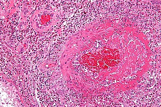
<span class="mw-page-title-main">Angioma</span> Medical condition

Angiomas are benign tumors derived from cells of the vascular or lymphatic vessel walls (endothelium) or derived from cells of the tissues surrounding these vessels.

<span class="mw-page-title-main">Fatty streak</span> Visible lesion indicating atherosclerosis

A fatty streak is the first grossly visible lesion in the development of atherosclerosis. It appears as an irregular yellow-white discoloration on the luminal surface of an artery. It consists of aggregates of foam cells, which are lipoprotein-loaded macrophages, located in the intima, the innermost layer of the artery, beneath the endothelial cells that layer the lumina through which blood flows. Fatty streaks may also include T cells, aggregated platelets, and smooth muscle cells. Although fatty streaks can develop into atheromas, not all are destined to become advanced lesions.

Colony-stimulating factors (CSFs) are secreted glycoproteins that bind to receptor proteins on the surfaces of committed progenitors in the bone marrow, thereby activating intracellular signaling pathways that can cause the cells to proliferate and differentiate into a specific kind of blood cell.

The uterine sarcomas form a group of malignant tumors that arises from the smooth muscle or connective tissue of the uterus.

<span class="mw-page-title-main">Primitive neuroectodermal tumor</span> Medical condition

Primitive neuroectodermal tumor is a malignant (cancerous) neural crest tumor. It is a rare tumor, usually occurring in children and young adults under 25 years of age. The overall 5 year survival rate is about 53%.

<span class="mw-page-title-main">Mixed Müllerian tumor</span> Medical condition

A malignant mixed Müllerian tumor, also known as malignant mixed mesodermal tumor (MMMT) is a cancer found in the uterus, the ovaries, the fallopian tubes and other parts of the body that contains both carcinomatous and sarcomatous components. It is divided into two types, homologous and a heterologous type. MMMT account for between two and five percent of all tumors derived from the body of the uterus, and are found predominantly in postmenopausal women with an average age of 66 years. Risk factors are similar to those of adenocarcinomas and include obesity, exogenous estrogen therapies, and nulliparity. Less well-understood but potential risk factors include tamoxifen therapy and pelvic irradiation.

<span class="mw-page-title-main">Medullary breast carcinoma</span> Rare type of breast cancer

Medullary breast carcinoma is a rare type of breast cancer that is characterized as a relatively circumscribed tumor with pushing, rather than infiltrating, margins. It is histologically characterized as poorly differentiated cells with abundant cytoplasm and pleomorphic high grade vesicular nuclei. It involves lymphocytic infiltration in and around the tumor and can appear to be brown in appearance with necrosis and hemorrhage. Prognosis is measured through staging but can often be treated successfully and has a better prognosis than other infiltrating breast carcinomas.

<span class="mw-page-title-main">Vinay Kumar (pathologist)</span> American pathologist

Vinay Kumar is the Lowell T. Coggeshall Distinguished Service Professor of Pathology at the University of Chicago, where he was also the Chairman (2000-2016) of the Department of Pathology. He is a recipient of Life Time Achievement Award by National Board of Examinations.

<span class="mw-page-title-main">Fat necrosis</span> Medical condition

Fat necrosis is a form of necrosis that is caused by the action of lipases on adipocytes.

Genital leiomyomas are leiomyomas that originate in the dartos muscles, or smooth muscles, of the genitalia, areola, and nipple. They are a subtype of cutaneous leiomyomas that affect smooth muscle found in the scrotum, labia, or nipple. They are benign tumors, but may cause pain and discomfort to patients. Genital leiomyoma can be symptomatic or asymptomatic and is dependent on the type of leiomyoma. In most cases, pain in the affected area or region is most common. For vaginal leiomyoma, vaginal bleeding and pain may occur. Uterine leiomyoma may exhibit pain in the area as well as painful bowel movement and/or sexual intercourse. Nipple pain, enlargement, and tenderness can be a symptom of nipple-areolar leiomyomas. Genital leiomyomas can be caused by multiple factors, one can be genetic mutations that affect hormones such as estrogen and progesterone. Moreover, risk factors to the development of genital leiomyomas include age, race, and gender. Ultrasound and imaging procedures are used to diagnose genital leiomyomas, while surgically removing the tumor is the most common treatment of these diseases. Case studies for nipple areolar, scrotal, and uterine leiomyoma were used, since there were not enough secondary resources to provide more evidence.

<span class="mw-page-title-main">Elliptocyte</span> Abnormal red blood cell

Elliptocytes, also known as ovalocytes or cigar cells, are abnormally shaped red blood cells that appear oval or elongated, from slightly egg-shaped to rod or pencil forms. They have normal central pallor with the hemoglobin appearing concentrated at the ends of the elongated cells when viewed through a light microscope. The ends of the cells are blunt and not sharp like sickle cells.

<span class="mw-page-title-main">Endometrial stromal sarcoma</span> Medical condition

Endometrial stromal sarcoma is a malignant subtype of endometrial stromal tumor arising from the stroma of the endometrium rather than the glands. There are three grades for endometrial stromal tumors, as follows. It was previously known as endolymphatic stromal myosis because of diffuse infiltration of myometrial tissue or the invasion of lymphatic channels.

Abul K. Abbas is an American pathologist at University of California San Francisco where he is Distinguished Professor in Pathology and former chair of its Department of Pathology. He is senior editor of the pathology reference book Robbins and Cotran Pathologic Basis of Disease along with Vinay Kumar, as well as Basic Immunology, and Cellular & Molecular Immunology. He was editor for Immunity from 1993 to 1996, and continues to serve as a member of the editorial board. He was one of the inaugural co-editors of the Annual Review of Pathology: Mechanisms of Disease for issues from 2006 to 2020. He has published nearly 200 scientific papers.

References

  1. 1 2 "Leiomyosarcoma - Overview - Mayo Clinic". www.mayoclinic.org. Retrieved 2023-10-04.
  2. 1 2 3 4 5 6 "Leiomyosarcoma - NCI". www.cancer.gov. 2020-06-22. Retrieved 2023-10-04.
  3. 1 2 "Basic info". Leiomyosarcoma.info. Archived from the original on 2009-04-30.
  4. 1 2 3 Kumar, Vinay; Abbas, Abul K.; Aster, Jon C. (2021). "Chapter 26: Bones, Joints, and Soft Tissue Tumors". Robbins & Cotran Pathologic Basis of Disease (10th ed.). Jeremy Bowes. p. 1208. ISBN   978-0-323-53113-9.
  5. Kumar, Vinay; Abbas, Abul K.; Aster, Jon C. (2021). "Chapter 26: Bones, Joints, and Soft Tissue Tumors". Robbins & Cotran Pathologic Basis of Disease (10th ed.). Jeremy Bowes. p.1213. ISBN   978-0-323-53113-9.
  6. Sue Ghosh; Jonathan Hecht; Tanaz Ferzandi; Christopher Awtrey (2007). "Leiomyosarcoma of the Uterus (ULMS): A Review". The Liddy Shriver Sarcoma Initiative. Retrieved 1 February 2016.
  7. Vijay Shankar, M.D. "Soft tissue - Smooth muscle - Leiomyosarcoma - general". Pathology Outlines. Topic Completed: 1 November 2012. Revised: 11 September 2019
  8. "About Chemotherapy". Leiomyosarcoma.info. August 2002. Archived from the original on 2009-06-25.
  9. Rastogi, Sameer; Bakhshi, Sameer (2016). "Trabectedin in Soft Tissue Sarcoma: Have We Hit the Bull's-eye?". Journal of Clinical Oncology. 34 (29): 3582–3583. doi:10.1200/jco.2015.65.7130. PMID   27458287.
  10. Rastogi, Sameer; Sharma, Aparna; Vanidassane, Ilavarasi; Aggarwal, Aditi; Mridha, Asitranjan; Pandey, Rambha; Dhamija, Ekta; Barwad, Adarsh (2019). "Pazopanib efficacy and toxicity in a metastatic sarcoma cohort: Are Indian patients different?". Indian Journal of Cancer. 56 (3): 207–210. doi: 10.4103/ijc.IJC_105_18 . PMID   31389382. S2CID   198168573.
  11. Abu-Rustum, N. R.; Curtin, J. P.; Burt, M.; Jones, W. B. (1997). "Regression of uterine low-grade smooth-muscle tumors metastatic to the lung after oophorectomy". Obstetrics and Gynecology. 89 (5 Pt 2): 850–852. doi:10.1016/S0029-7844(97)00033-1. PMID   9166348. S2CID   23049718.
  12. Hardman, M. P.; Roman, J. J.; Burnett, A. F.; Santin, A. D. (2007). "Metastatic Uterine Leiomyosarcoma Regression Using an Aromatase Inhibitor". Obstetrics & Gynecology. 110 (2 Pt 2): 518–520. doi:10.1097/01.AOG.0000267533.56546.c2. PMID   17666649. S2CID   24257210.
  13. "Uterine Sarcoma Clinical Trials". National Cancer Institute.
  14. Kumar, Vinay; Abbas, Abul K.; Aster, Jon C. (2021). "Chapter 26: Bones, Joints, and Soft Tissue Tumors". Robbins & Cotran Pathologic Basis of Disease (10th ed.). Jeremy Bowes. p.1014. ISBN   978-0-323-53113-9
  15. 1 2 3 Sun, S.; Bonaffini, P. A.; Nougaret, S.; Fournier, L.; Dohan, A.; Chong, J.; Smith, J.; Addley, H.; Reinhold, C. (October 2019). "How to differentiate uterine leiomyosarcoma from leiomyoma with imaging". Diagnostic and Interventional Imaging. 100 (10): 619–634. doi:10.1016/j.diii.2019.07.007. ISSN   2211-5684. PMID   31427216.
  16. Kumar, Vinay; Abbas, Abul K.; Aster, Jon C. (2021). "Chapter 26: Bones, Joints, and Soft Tissue Tumors". Robbins & Cotran Pathologic Basis of Disease (10th ed.). Jeremy Bowes. p.1015. ISBN   978-0-323-53113-9
  17. Health, Center for Devices and Radiological (2020-12-30). "UPDATE: Perform Only Contained Morcellation When Laparoscopic Power Morcellation Is Appropriate: FDA Safety Communication". FDA.
  18. "Leicester legend Weller mourned". BBC Online. 2004-11-13. Retrieved 2010-05-14.
  19. "Jordan treated for cancer". BBC Online. 2002-08-11. Retrieved 2009-10-30.
  20. Gillespie, Kerry (2008-04-12). "'Can't ever give up hope,' Basrur says". The Toronto Star . Retrieved 2010-01-08.
  21. Brown, Stacia. "Diana Sands: What Was and What Could've Been". Archived from the original on 6 December 2017. Retrieved 25 May 2017.
  22. "That's My Time". Irwinbarker.com. Archived from the original on 2008-10-02. Retrieved 5 March 2015. In Irwin's own words: 'Cancer has my body but not my spirit, and I'll continue to make jokes, not so much about cancer, but in spite of it.'
  23. Fiona Hackett (2009-11-27). "Former player Linda Uttley". waspsfc.co.uk. Retrieved 2022-02-28.