Related Research Articles

Medical ultrasound is a diagnostic imaging technique, or therapeutic application of ultrasound. It is used to create an image of internal body structures such as tendons, muscles, joints, blood vessels, and internal organs. Its aim is often to find a source of a disease or to exclude pathology. The practice of examining pregnant women using ultrasound is called obstetric ultrasound, and was an early development and application of clinical ultrasonography.

Adenocarcinoma (AC) is a type of cancerous tumor that can occur in several parts of the body. It is defined as neoplasia of epithelial tissue that has glandular origin, glandular characteristics, or both. Adenocarcinomas are part of the larger grouping of carcinomas, but are also sometimes called by more precise terms omitting the word, where these exist. Thus invasive ductal carcinoma, the most common form of breast cancer, is adenocarcinoma but does not use the term in its name—however, esophageal adenocarcinoma does to distinguish it from the other common type of esophageal cancer, esophageal squamous cell carcinoma. Several of the most common forms of cancer are adenocarcinomas, and the various sorts of adenocarcinoma vary greatly in all their aspects, so that few useful generalizations can be made about them.

Alpha-fetoprotein is a protein that in humans is encoded by the AFP gene. The AFP gene is located on the q arm of chromosome 4 (4q25). Maternal AFP serum level is used to screen for Down syndrome, neural tube defects, and other chromosomal abnormalities.

A teratoma is a tumor made up of several different types of tissue, such as hair, muscle, teeth, or bone. Teratomata typically form in the ovary, testicle, or coccyx.

A lipoma is a benign tumor made of fat tissue. They are generally soft to the touch, movable, and painless. They usually occur just under the skin, but occasionally may be deeper. Most are less than 5 cm in size. Common locations include upper back, shoulders, and abdomen. It is possible to have a number of lipomas.
Spinal tumors are neoplasms located in either the vertebral column or the spinal cord. There are three main types of spinal tumors classified based on their location: extradural and intradural. Extradural tumors are located outside the dura mater lining and are most commonly metastatic. Intradural tumors are located inside the dura mater lining and are further subdivided into intramedullary and extramedullary tumors. Intradural-intramedullary tumors are located within the dura and spinal cord parenchyma, while intradural-extramedullary tumors are located within the dura but outside the spinal cord parenchyma. The most common presenting symptom of spinal tumors is nocturnal back pain. Other common symptoms include muscle weakness, sensory loss, and difficulty walking. Loss of bowel and bladder control may occur during the later stages of the disease.

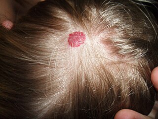
Dermatofibrosarcoma protuberans (DFSP) is a rare locally aggressive malignant cutaneous soft-tissue sarcoma. DFSP develops in the connective tissue cells in the middle layer of the skin (dermis). Estimates of the overall occurrence of DFSP in the United States are 0.8 to 4.5 cases per million persons per year. In the United States, DFSP accounts for between 1 and 6 percent of all soft tissue sarcomas and 18 percent of all cutaneous soft tissue sarcomas. In the Surveillance, Epidemiology and End Results (SEER) tumor registry from 1992 through 2004, DFSP was second only to Kaposi sarcoma.

A benign tumor is a mass of cells (tumor) that lacks the ability to either invade neighboring tissue or metastasize. When removed, benign tumors usually do not grow back, whereas malignant tumors sometimes do. Unlike most benign tumors elsewhere in the body, benign brain tumors can be life-threatening. Benign tumors generally have a slower growth rate than malignant tumors and the tumor cells are usually more differentiated. They are typically surrounded by an outer surface or stay contained within the epithelium. Common examples of benign tumors include moles and uterine fibroids.

Fibroadenomas are benign breast tumours characterized by an admixture of stromal and epithelial tissue. Breasts are made of lobules and ducts. These are surrounded by glandular, fibrous and fatty tissues. Fibroadenomas develop from the lobules. The glandular tissue and ducts grow over the lobule to form a solid lump.

A hamartoma is a mostly benign, local malformation of cells that resembles a neoplasm of local tissue but is usually due to an overgrowth of multiple aberrant cells, with a basis in a systemic genetic condition, rather than a growth descended from a single mutated cell (monoclonality), as would typically define a benign neoplasm/tumor. Despite this, many hamartomas are found to have clonal chromosomal aberrations that are acquired through somatic mutations, and on this basis the term hamartoma is sometimes considered synonymous with neoplasm. Hamartomas are by definition benign, slow-growing or self-limiting, though the underlying condition may still predispose the individual towards malignancies.

Hemangioendotheliomas are a family of vascular neoplasms of intermediate malignancy.

High-intensity focused ultrasound (HIFU) is a non-invasive therapeutic technique that uses non-ionizing ultrasonic waves to heat or ablate tissue. HIFU can be used to increase the flow of blood or lymph, or to destroy tissue, such as tumors, via thermal and mechanical mechanisms. Given the prevalence and relatively low cost of ultrasound, HIFU has been subject to much research and development. The premise of HIFU is that it is a non-invasive low cost therapy that can at minimum outperform the current standard of care.
Sacrococcygeal teratoma (SCT) is a type of tumor known as a teratoma that develops at the base of the coccyx (tailbone) and is thought to be derived from the primitive streak. Sacrococcygeal teratomas are benign 75% of the time, malignant 12% of the time, and the remainder are considered "immature teratomas" that share benign and malignant features. Benign sacrococcygeal teratomas are more likely to develop in younger children who are less than 5 months old, and older children are more likely to develop malignant sacrococcygeal teratomas. The Currarino triad, due to an autosomal dominant mutation in the MNX1 gene, consists of a presacral mass, anorectal malformation and sacral dysgenesis.
A vascular tumor is a tumor of vascular origin; a soft tissue growth that can be either benign or malignant, formed from blood vessels or lymph vessels. Examples of vascular tumors include hemangiomas, lymphangiomas, hemangioendotheliomas, Kaposi's sarcomas, angiosarcomas, and hemangioblastomas. An angioma refers to any type of benign vascular tumor.
A hibernoma is a benign neoplasm of vestigial brown fat. The term was originally used by the French anatomist Louis Gery in 1914.

Melanotic neuroectodermal tumor of infancy is a very rare oral cavity tumor that is seen in patients usually at or around birth. It must be removed to be cured. Definitions: A rare, biphasic, neuroblastic, and pigmented epithelial neoplasm of craniofacial sites, usually involving the oral cavity or gums.

Endodermal sinus tumor (EST) is a member of the germ cell tumor group of cancers. It is the most common testicular tumor in children under 3, and is also known as infantile embryonal carcinoma. This age group has a very good prognosis. In contrast to the pure form typical of infants, adult endodermal sinus tumors are often found in combination with other kinds of germ cell tumor, particularly teratoma and embryonal carcinoma. While pure teratoma is usually benign, endodermal sinus tumor is malignant.

Vaginal cysts are uncommon benign cysts that develop in the vaginal wall. The type of epithelial tissue lining a cyst is used to classify these growths. They can be congenital. They can present in childhood and adulthood. The most common type is the squamous inclusion cyst. It develops within vaginal tissue present at the site of an episiotomy or other vaginal surgical sites. In most instances they do not cause symptoms and present with few or no complications. A vaginal cyst can develop on the surface of the vaginal epithelium or in deeper layers. Often, they are found by the women herself and as an incidental finding during a routine pelvic examination. Vaginal cysts can mimic other structures that protrude from the vagina such as a rectocele and cystocele. Some cysts can be distinguished visually but most will need a biopsy to determine the type. Vaginal cysts can vary in size and can grow as large as 7 cm. Other cysts can be present on the vaginal wall though mostly these can be differentiated. Vaginal cysts can often be palpated (felt) by a clinician. Vaginal cysts are one type of vaginal mass, others include cancers and tumors. The prevalence of vaginal cysts is uncertain since many go unreported but it is estimated that 1 out of 200 women have a vaginal cyst. Vaginal cysts may initially be discovered during pregnancy and childbirth. These are then treated to provide an unobstructed delivery of the infant. Growths that originate from the urethra and other tissue can present as cysts of the vagina.
Epignathus is a rare teratoma of the oropharynx. Epignathus is a form of oropharyngeal teratoma that arises from the palate and, in most cases, results in death. The pathology is thought to be due to unorganized and uncontrolled differentiation of somatic cells leading to formation of the teratoma; sometimes it is also referred to as "fetus-in-fetu", which is an extremely rare occurrence of an incomplete but parasitic fetus located in the body of its twin. This tumor is considered benign but life-threatening because of its atypical features and high risk of airway obstruction, which is the cause of death in 80-100% of the cases at the time of delivery.
References
- ↑ Rapini, Ronald P.; Bolognia, Jean L.; Jorizzo, Joseph L. (2007). Dermatology: 2-Volume Set. St. Louis: Mosby. ISBN 978-1-4160-2999-1.
- ↑ Kumar, A.; Brierley, D.; Hunter, K.D.; Lee, N. (November 2015). "Rapidly-growing buccal mass in a 6-month-old infant". British Journal of Oral and Maxillofacial Surgery. 53 (9): 888–890. doi:10.1016/j.bjoms.2015.07.006. PMID 26250364.
- ↑ James, William; Berger, Timothy; Elston, Dirk (2005). Andrews' Diseases of the Skin: Clinical Dermatology. (10th ed.). Saunders. ISBN 0-7216-2921-0.
- ↑ Vellios, F; Baez, J; Shumacker, HB (November 1958). "Lipoblastomatosis: a tumor of fetal fat different from hibernoma; report of a case, with observations on the embryogenesis of human adipose tissue". The American Journal of Pathology . 34 (6): 1149–59. PMC 1934796 . PMID 13583102.
- ↑ Gammelgaard, Niels; Jørgensen, Karstein; Lund, Claus (July 1983). "Benign Lipoblastoma in the Neck Causing Respiratory Insufficiency". The Laryngoscope . 93 (7): 935–937. doi:10.1288/00005537-198307000-00017. PMID 6865630. S2CID 35331275.
- ↑ Robinson, Philip; Vanhoenacker, Filip M. (2017). "Adipocytic Tumors". Imaging of Soft Tissue Tumors: 197–241. doi:10.1007/978-3-319-46679-8_12. ISBN 978-3-319-46677-4.
- ↑ Ahlawat, Shivani; M. Fayad, Laura (2020). "Revisiting the WHO classification system of soft tissue tumours: emphasis on advanced magnetic resonance imaging sequences. Part 1". Polish Journal of Radiology. 85 (1): 396–408. doi:10.5114/pjr.2020.98685. PMC 7509695 . PMID 32999693.
- 1 2 3 Kamal, Achmad Fauzi; Wiratnaya, I. Gde Eka; Hutagalung, Errol Untung; Prasetyo, Marcel; Kodrat, Evelina; Widodo, Wahyu; Effendi, Zuhri; Husodo, Kurniadi (2014-09-15). "Lipoblastoma and Lipoblastomatosis of the Lower Leg". Case Reports in Orthopedics. 2014: 582876. doi:10.1155/2014/582876. PMC 4181785 . PMID 25302126.
- 1 2 Calobrisi, Stella D.; Garland, Jeffery S.; Esterly, Nancy B. (1998). "Congenital Lipoblastomatosis of the Lower Extremity in a Neonate". Pediatric Dermatology. 15 (3): 210–213. doi:10.1111/j.1525-1470.1998.tb01318.x. PMID 9655318. S2CID 45439781.