| Histiocytic sarcoma | |
|---|---|
| Specialty | Oncology, angiology |
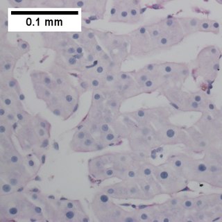
Histiocytic sarcoma is a tumor derived from histiocytes. [1] The tumor is often positive for CD163 [2] and can appear in the thyroid. [3] However, in some cases it can also appear in the brain.
| Histiocytic sarcoma | |
|---|---|
| Specialty | Oncology, angiology |
Histiocytic sarcoma is a tumor derived from histiocytes. [1] The tumor is often positive for CD163 [2] and can appear in the thyroid. [3] However, in some cases it can also appear in the brain.

Gastrointestinal stromal tumors (GISTs) are the most common mesenchymal neoplasms of the gastrointestinal tract. GISTs arise in the smooth muscle pacemaker interstitial cell of Cajal, or similar cells. They are defined as tumors whose behavior is driven by mutations in the KIT gene (85%), PDGFRA gene (10%), or BRAF kinase (rare). 95% of GISTs stain positively for KIT (CD117). Most (66%) occur in the stomach and gastric GISTs have a lower malignant potential than tumors found elsewhere in the GI tract.

Tumors of the hematopoietic and lymphoid tissues or tumours of the haematopoietic and lymphoid malignancies are tumors that affect the blood, bone marrow, lymph, and lymphatic system. Because these tissues are all intimately connected through both the circulatory system and the immune system, a disease affecting one will often affect the others as well, making myeloproliferation and lymphoproliferation closely related and often overlapping problems.

Dermatofibrosarcoma protuberans (DFSP) is a rare tumor of the dermis layer of the skin, and is classified as a sarcoma. There is only about one case per million per year. DFSP is a fibrosarcoma, more precisely a cutaneous soft tissue sarcoma. In many respects, the disease behaves as a benign tumor, but in 2–5% of cases it can metastasize, so it should be considered to have malignant potential. It occurs most often in adults in their thirties; it has been described congenitally, in children, and the elderly. It accounts for approximately 2–6% of soft tissue sarcoma cancers.
![Myeloid sarcoma solid tumor composed of immature white blood cells[2] called myeloblasts.](https://upload.wikimedia.org/wikipedia/commons/thumb/1/18/Chloroma_-_very_high_mag.jpg/320px-Chloroma_-_very_high_mag.jpg)
A myeloid sarcoma, is a solid tumor composed of immature white blood cells called myeloblasts. A chloroma is an extramedullary manifestation of acute myeloid leukemia; in other words, it is a solid collection of leukemic cells occurring outside of the bone marrow.

Leiomyosarcoma, is a malignant (cancerous) smooth muscle tumor. A benign tumor originating from the same tissue is termed leiomyoma. While it has been believed that leiomyosarcomas do not arise from leiomyomas, there are leiomyoma variants for which classification is evolving.

Giant-cell tumor of the bone (GCTOB), is a relatively uncommon tumor of the bone. It is characterized by the presence of multinucleated giant cells. Malignancy in giant-cell tumor is uncommon and occurs in about 2% of all cases. However, if malignant degeneration does occur, it is likely to metastasize to the lungs. Giant-cell tumors are normally benign, with unpredictable behavior. It is a heterogeneous tumor composed of three different cell populations. The giant-cell tumour stromal cells (GCTSC) constitute the neoplastic cells, which are from an osteoblastic origin and are classified based on expression of osteoblast cell markers such as alkaline phosphatase and osteocalcin. In contrast, the mononuclear histiocytic cells (MNHC) and multinucleated giant cell (MNGC) fractions are secondarily recruited and comprise the non-neoplastic cell population. They are derived from an osteoclast-monocyte lineage determined primarily by expression of CD68, a marker for monocytic precursor cells. In most patients, the tumors are slow to develop, but may recur locally in as many as 50% of cases.

Platelet endothelial cell adhesion molecule (PECAM-1) also known as cluster of differentiation 31 (CD31) is a protein that in humans is encoded by the PECAM1 gene found on chromosome17q23.3. PECAM-1 plays a key role in removing aged neutrophils from the body.
Primary effusion lymphoma (PEL) is a B-cell lymphoma, presenting with a malignant effusion without a tumor mass.
Malignant histiocytosis is a rare hereditary disease found in the Bernese Mountain Dog and humans, characterized by histiocytic infiltration of the lungs and lymph nodes. The liver, spleen, and central nervous system can also be affected. Histiocytes are a component of the immune system that proliferate abnormally in this disease. In addition to its importance in veterinary medicine, the condition is also important in human pathology.

A histiocytoma in the dog is a benign tumor. It is an abnormal growth in the skin of histiocytes (histiocytosis), a cell that is part of the immune system. A similar disease in humans, Hashimoto-Pritzker disease, is also a Langerhans cell histiocytosis. Dog breeds that may be more at risk for this tumor include Bulldogs, American Pit Bull Terriers, American Staffordshire Terriers, Scottish Terriers, Greyhounds, Boxers, and Boston Terriers. They also rarely occur in goats and cattle.

Erdheim–Chester disease is a rare disease characterized by the abnormal multiplication of a specific type of white blood cells called histiocytes, or tissue macrophages. It was declared a histiocytic neoplasm by the World Health Organization in 2016. Onset typically is in middle age. The disease involves an infiltration of lipid-laden macrophages, multinucleated giant cells, an inflammatory infiltrate of lymphocytes and histiocytes in the bone marrow, and a generalized sclerosis of the long bones.

Ewing's sarcoma is a type of cancer that forms in bone or soft tissue. Symptoms may include swelling and pain at the site of the tumor, fever, and a bone fracture. The most common areas where it begins are the legs, pelvis, and chest wall. In about 25% of cases, the cancer has already spread to other parts of the body at the time of diagnosis. Complications may include a pleural effusion or paraplegia.

Rosai–Dorfman disease, also known as sinus histiocytosis with massive lymphadenopathy or sometimes as Destombes–Rosai–Dorfman disease, is a rare disorder of unknown cause that is characterized by abundant histiocytes in the lymph nodes or other locations throughout the body.

Cluster of differentiation 97 is a protein also known as BL-Ac[F2] encoded by the ADGRE5 gene. CD97 is a member of the adhesion G protein-coupled receptor (GPCR) family. Adhesion GPCRs are characterized by an extended extracellular region often possessing N-terminal protein modules that is linked to a TM7 region via a domain known as the GPCR-Autoproteolysis INducing (GAIN) domain.

Epithelioid sarcoma is a rare soft tissue sarcoma arising from mesenchymal tissue and characterized by epithelioid-like features. It accounts for less than 1% of all soft tissue sarcomas. It was first clearly characterized by F.M. Enzinger in 1970. It commonly presents itself in the distal limbs of young adults as a small, soft mass or a series of bumps. A proximal version has also been described, frequently occurring in the upper extremities. Rare cases have been reported in the pelvis, vulva, penis, and spine.

Kaposi's sarcoma (KS) is a type of cancer that can form masses in the skin, lymph nodes, or other organs. The skin lesions are usually purple in color. They can occur singularly, in a limited area, or be widespread. It may worsen either gradually or quickly. Lesions may be flat or raised. Human herpesvirus 8 (HHV8) is found in the lesions of all those who are affected. Risk factors include poor immune function, either as a result of disease or specific medications, and chronic lymphedema.

Follicular dendritic cell sarcoma (FDCS) is an extremely rare neoplasm. While the existence of FDC tumors was predicted by Lennert in 1978, the tumor wasn't fully recognized as its own cancer until 1986 after characterization by Monda et al. It accounts for only 0.4% of soft tissue sarcomas, but has significant recurrent and metastatic potential and is considered an intermediate grade malignancy. The major hurdle in treating FDCS has been misdiagnosis. It is a newly characterized cancer, and because of its similarities in presentation and markers to lymphoma, both Hodgkin and Non-Hodgkin subtypes, diagnosis of FDCS can be difficult. With recent advancements in cancer biology better diagnostic assays and chemotherapeutic agents have been made to more accurately diagnose and treat FDCS.
Histiocytic diseases in dogs are a group of diseases in dogs which may involve the skin, and which can be difficult to differentiate from granulomatous, reactive inflammatory or lymphoproliferative diseases. The clinical presentation and behaviour as well as response to therapy vary greatly among the syndromes.
The Xanthogranulomatous Process (XP), is a form of acute and chronic inflammation characterized by an exuberant clustering of foamy macrophages among other inflammatory cells. Localization in the kidney and renal pelvis has been the most frequent and better known occurrence followed by that in the gallbladder but many others have been subsequently recorded. The pathological findings of the process and etiopathogenetic and clinical observations have been reviewed by Cozzutto and Carbone.
T-cell/histiocyte-rich large B-cell lymphoma (THRLBCL) is a malignancy of B-cells. B-cells are lymphocytes that normally function in the humoral immunity component of the adaptive immune system by secreting antibodies that, for example, bind to and neutralize invasive pathogens. Among the various forms of B-cell lymphomas, THRLBCL is a rarely occurring subtype of the diffuse large B-cell lymphomas (DLBCL). DLBCL are a large group of lymphomas that account for ~25% of all non-Hodgkin lymphomas worldwide. THRLBCL is distinguished from the other DLBCL subtypes by the predominance of non-malignant T-cell lymphocytes and histiocytes over malignant B-cells in its tumors and tissue infiltrates.
| Classification | |
|---|---|
| External resources |
| This article about a neoplasm is a stub. You can help Wikipedia by expanding it. |