There are inherited (primary HLH) and acquired (secondary HLH) forms. The inherited form is due to genetic mutations and usually presents in infants and children, with a median age of onset of 3–6 months.[2] Familial HLH is an autosomal recessive disease, hence each sibling of a child with familial HLH has a 25% chance of developing the disease, a 50% chance of carrying the defective gene (which is very rarely associated with any risk of disease), and a 25% chance of not being affected and not carrying the gene defect.[3] Genes that are commonly mutated in those with primary HLH lead to defective lymphocyte (natural killer cell and cytotoxic T-cell) function. The mutated genes are PRF1 (perforin-1), UNC13D, STX11, and STXBP2.[2] Secondary HLH usually presents in adulthood (usually in people with genetic changes predisposing them to the disease) after exposure to a trigger. Common triggers leading to secondary HLH include infections, cancer, or autoimmune diseases.[2] The incidence of all forms of HLH was estimated to be 4.2 cases per 1 million people in a population based study from England in 2018, but the true incidence is not known.[4] The incidence of HLH (especially secondary HLH) is thought to be underestimated as the clinical signs and symptoms are very similar to sepsis.[2]
The onset of HLH occurs before the age of one year in approximately 70% of cases. Familial HLH should be suspected if siblings are diagnosed with HLH or if symptoms recur when therapy has been stopped.[3]
Five genetic subtypes (FHL1, FHL2, FHL3, FHL4, and FHL5) are described, with an estimated overall prevalence of one in 50,000 and equal gender distribution. Molecular genetic testing for four of the causative genes, PRF1 (FHL2), UNC13D (FHL3), STX11 (FHL4), and STXBP2 (FHL5), is available on a clinical basis. Symptoms of FHL are usually evident within the first few months of life and may even develop in utero. However, symptomatic presentation throughout childhood and even into young adulthood has been observed in some cases.[citation needed]
The five subtypes of FHL[14] are each associated with a specific gene:
Nearly half of the cases of type 2 familial hemophagocytic lymphohistiocytosis are due to bi-allelic PRF1 mutations.[15]
Pathophysiology
The underlying causes, either inherited or acquired, lead to an unchecked immune response when exposed to triggers. Impaired NK-cell cytotoxicity is the hallmark of HLH. All genetic defects for familial HLH are related to granule-dependent cytotoxicity. This inability to remove infected and antigen-presenting cells and terminate the immune response leads to uncontrolled proliferation and activation of the immune system with the release of excessive cytokines. These cells then infiltrate organs, releasing more cytokines, which gives the clinical picture. The fever is caused by IL-1, IL-6 and TNF-alpha; the cytopenia is due to the suppressive effect on hematopoiesis by TNF-alpha and TNF-gamma. TNF-alpha and TNF-gamma may also lead to inhibition of lipoprotein lipase or stimulate triglyceride synthesis. Activated macrophages secrete ferritin and plasminogen activator leading to hyperfibrinolysis.[16]
The serum C reactive protein, erythrocyte sedimentation rate, and ferritin levels are markedly elevated. In children, ferritin levels above 10000 μg/L are very sensitive and specific for the diagnosis of HLH,[17] however, the diagnostic utility for ferritin is less for adult HLH patients.[18]
Primary HLH, also known as familial haemophagocytic lymphohistiocytosis (FHL) or familial erythrophagocytic lymphohistiocytosis, is a heterogeneous autosomal recessive disorder found to be more prevalent with parental consanguinity.[citation needed]
Secondary haemophagocytic lymphohistiocytosis (acquired haemophagocytic lymphohistiocytosis) occurs after strong immunologic activation, such as that which can occur with systemic infection, immunodeficiency, or underlying malignancy.[21]
Both forms are characterized by the overwhelming activation of normal T lymphocytes and macrophages, invariably leading to clinical and haematologic alterations and death in the absence of treatment.[citation needed]
An atypical presentation of primary HLH where the inflammation is limited to the central nervous system has been described.[22][23]
Diagnostic criteria
The current (2004) diagnostic criteria for HLH are:[24][2]
1. A molecular diagnosis consistent with HLH. These include the identification of pathologic mutations of PRF1, UNC13D, or STX11.
OR
2. Fulfillment of five out of the eight criteria below:
Soluble CD25 (soluble IL-2 receptor) >2400 U/ml (or per local reference laboratory)
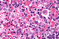
Despite the disorder's name, hemophagocytosis is not required to be present for the diagnosis. It may not be present in early stages of the disordered inflammation.[2]
Not all five out of eight criteria are required for diagnosis of HLH in adults, and a high index of suspicion is required for diagnosis as delay results in increased mortality. The diagnostic criteria were developed in pediatric populations and have not been validated for adult HLH patients.[25] Attempts to improve diagnosis of HLH have included use of the HScore, which can be used to estimate an individual's risk of HLH.[26] In adults, soluble IL-2 receptor has been found to be a very sensitive marker for HLH, demonstrating 100% sensitivity for ruling out HLH below a cutoff of 2400 U/mL and optimal cutoff for ruling in at 2515 U/mL (sensitivity, 100%; specificity, 72.5%), with 93% specificity at >10 000 U/mL.[27]
Other conditions that may be confused with this condition include autoimmune lymphoproliferative syndrome.[28] As a syndrome of intense inflammation it needs to be differentiated from sepsis, which may be extremely challenging.[29]
The diagnosis of acquired, or secondary, HLH is usually made in association with infection by viruses, bacteria, fungi, or parasites or in association with lymphoma, autoimmune disease, or metabolic disease. Acquired HLH may have decreased, normal, or increased NK cell activity.[citation needed]
Griscelli syndrome
A major differential diagnosis of HLH is Griscelli syndrome (type 2). This is a rare autosomal recessive disorder characterized by partial albinism, hepatosplenomegaly, pancytopenia, hepatitis, immunologic abnormalities, and lymphohistiocytosis. Most cases have been diagnosed between 4 months and 7 years of age, with a mean age of about 17 months.[citation needed]
Three types of Griscelli syndrome are recognised: type 1 has neurologic symptoms and mutations in MYO5A. The prognosis depends on the severity of neurologic manifestations. Type 2 has mutations in RAB27A and haemophagocytic syndrome, with abnormal T-cell and macrophage activation. This type has a grave prognosis if untreated. Type 3 has mutations in melanophilin and is characterized by partial albinism. This type does not pose a threat to those so affected.[citation needed]
Treatment
HLH is a description of an immunophysiologic state in time. It can be dangerous to infer a genetic impairment of granule-mediated cytotoxicity in patients, especially older children and adults, who meet any of the various criteria for HLH. Thus, like shock, one must simultaneously manage both the acute physiologic changes associated with HLH (like systemic inflammation, DIC, hepatitis, etc.) and look deeply for various underlying contributors.[citation needed]
The International Histiocyte Society has collected the published consensus management documents for the many contexts in which HLH occurs and they host full-text versions.[30]
Most patients who meet HLH criteria will have secondary cases. Treatment for these patients should focus on the underlying contributors. Additionally, treatment of the inflammation of HLH itself is often required.[citation needed]
While optimal treatment of HLH is still being debated, current treatment regimes usually involve high dose corticosteroids, etoposide and cyclosporin.[citation needed] Intravenous immunoglobulin is also used. Methotrexate and vincristine have also been used. However, for secondary HLH, evidence suggests that the benefit of etoposide-based therapy is limited, as it may mask or delay diagnosis of the underlying contributors and increase risk of toxicity. Cytokine targeted therapy, which is less toxic, could be a more rational approach to treat inflammation while not interfering with the identification of underlying causes of secondary HLH.[31]
On 20 November 2018, the FDA approved the anti-IFN-gamma monoclonal antibody emapalumab (proprietary name Gamifant) for the treatment of pediatric and adult primary HLH.[32]
In October 2021 NHS England published Clinical Commissioning Policy: Anakinra for Haemophagocytic Lymphohistiocytosis (HLH) for adults and children in all ages, allowing Anakinra (a modified recombinant interleukin 1 receptor antagonist) to be used in the treatment of HLH.[33]
People with HLH, especially when untreated, may need intensive therapy. Therefore, HLH should be included in the differential diagnosis of intensive care unit patients with cytopenia and hyperferritinemia.[34] Patients in the earlier stages of HLH are frequently hospitalized at internal medicine wards.[35]
Prognosis
The prognosis is guarded with an overall mortality of 50%. Poor prognostic factors included HLH associated with malignancy, with half the patients dying by 1.4 months compared to 22.8 months for non-tumour associated HLH patients.[36]
Secondary HLH in some individuals may be self-limited because patients can fully recover after having received only supportive medical treatment (i.e., IV immunoglobulin only). However, long-term remission without the use of cytotoxic and immune-suppressive therapies is unlikely in the majority of adults with HLH and in those with involvement of the central nervous system (brain and/or spinal cord).[14]
History
The first case report of HLH was published in 1939 under the term "Histiocytic Medullary Reticulosis".[37] A second report would come out in 1952 that would rename the disorder that same year.[38] Development of higher immune effector cell-associated hemophagocytic lymphohistiocytsis-like syndrome (IEC-HS) was observed in phase 1 clinical trails of the therapeutic CRG-023 developed by Cargo Therapeutics. In this study 18% of participants developed IEC-HS and an undisclosed number of patients died.[39]
Research
A 2018 systematic review of dengue-associated HLH cases reported pooled proportions in presentations of fever 97.2%, hepatomegaly 70.2%, splenomegaly 78.4%, thrombocytopenia 90.1%, anemia 76.0%, and serum ferritin ≥500 μg/L 97.1%. The case fatality rate was 14.6% among dengue HLH patients.[6]
12345678910111213Henter, Jan-Inge (6 February 2025). "Hemophagocytic Lymphohistiocytosis". New England Journal of Medicine. 392 (6): 584–598. doi:10.1056/NEJMra2314005.
12Esteban, Ysabella M.; de Jong, Jill L. O.; Tesher, Melissa S. (1 August 2017). "An Overview of Hemophagocytic Lymphohistiocytosis". Pediatric Annals. 46 (8): e309–13. doi:10.3928/19382359-20170717-01. PMID28806468.
↑Machowicz R, Janka G, Wiktor-Jedrzejczak W (March 2017). "Similar but not the same: Differential diagnosis of HLH and sepsis". Critical Reviews in Oncology/Hematology. 114: 1–12. doi:10.1016/j.critrevonc.2017.03.023. PMID28477737.
This page is based on this Wikipedia article Text is available under the CC BY-SA 4.0 license; additional terms may apply. Images, videos and audio are available under their respective licenses.