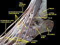
Extensor digitorum longus muscle

Last updated
Extensor digitorum longus muscle
Gray1241.png
The mucous sheaths of the tendons around the ankle. Lateral aspect. (Extensor dig. longus labeled at upper right.)
Extensor digitorum longus muscle - animation 2.gif
Animation
Details
Origin Anterior lateral condyle of tibia, anterior shaft of fibula and superior 34 of interosseous membrane
Insertion Dorsal surface; middle and distal phalanges of lateral four digits
Artery anterior tibial artery
Nerve deep fibular nerve
Actions extension of toes and dorsiflexion of ankle
Antagonist Flexor digitorum longus, Flexor digitorum brevis
Identifiers
Latin musculus extensor digitorum longus
TA98 A04.7.02.038
TA2 2645
FMA 22534
Anatomical terms of muscle

The extensor digitorum longus is a pennate muscle, situated at the lateral part of the front of the leg.

Contents

Structure

It arises from the lateral condyle of the tibia; from the upper three-quarters of the anterior surface of the body of the fibula; from the upper part of the interosseous membrane; from the deep surface of the fascia; and from the intermuscular septa between it and the tibialis anterior on the medial, and the peroneal muscles on the lateral side. Between it and the tibialis anterior are the upper portions of the anterior tibial vessels and deep peroneal nerve.

The muscle passes under the superior and inferior extensor retinaculum of foot in company with the fibularis tertius, and divides into four slips, which run forward on the dorsum of the foot, and are inserted into the second and third phalanges of the four lesser toes.

The tendons to the second, third, and fourth toes are each joined, opposite the metatarsophalangeal articulations, on the lateral side by a tendon of the extensor digitorum brevis. The tendons are inserted in the following manner: each receives a fibrous expansion from the interossei and lumbricals, and then spreads out into a broad aponeurosis, which covers the dorsal surface of the first phalanx: this aponeurosis, at the articulation of the first with the second phalanx, divides into three slips—an intermediate, which is inserted into the base of the second phalanx; and two collateral slips, which, after uniting on the dorsal surface of the second phalanx, are continued onward, to be inserted into the base of the third phalanx.

Variations

This muscle varies considerably in the modes of origin and the arrangement of its various tendons.

The tendons to the second and fifth toes may be found doubled, or extra slips are given off from one or more tendons to their corresponding metatarsal bones, or to the short extensor, or to one of the interosseous muscles.

A slip to the great toe from the innermost tendon has been found.

See also

Additional Images

Related Research Articles

<span class="mw-page-title-main">Foot</span> Anatomical structure found in vertebrates

The foot is an anatomical structure found in many vertebrates. It is the terminal portion of a limb which bears weight and allows locomotion. In many animals with feet, the foot is a separate organ at the terminal part of the leg made up of one or more segments or bones, generally including claws and or nails.

<span class="mw-page-title-main">Human leg</span> Lower extremity or limb of the human body (foot, lower leg, thigh and hip)

The human leg, in the general word sense, is the entire lower limb of the human body, including the foot, thigh or sometimes even the hip or gluteal region. However, the definition in human anatomy refers only to the section of the lower limb extending from the knee to the ankle, also known as the crus or, especially in non-technical use, the shank. Legs are used for standing, and all forms of locomotion including recreational such as dancing, and constitute a significant portion of a person's mass. Female legs generally have greater hip anteversion and tibiofemoral angles, but shorter femur and tibial lengths than those in males.

<span class="mw-page-title-main">Cuboid bone</span> Bone of the ankle

In the human body, the cuboid bone is one of the seven tarsal bones of the foot.

<span class="mw-page-title-main">Tibia</span> Shin bone

The tibia, also known as the shinbone or shankbone, is the larger, stronger, and anterior (frontal) of the two bones in the leg below the knee in vertebrates ; it connects the knee with the ankle. The tibia is found on the medial side of the leg next to the fibula and closer to the median plane. The tibia is connected to the fibula by the interosseous membrane of leg, forming a type of fibrous joint called a syndesmosis with very little movement. The tibia is named for the flute tibia. It is the second largest bone in the human body, after the femur. The leg bones are the strongest long bones as they support the rest of the body.

<span class="mw-page-title-main">Fibula</span> Leg bone on the lateral side of the tibia

The fibula or calf bone is a leg bone on the lateral side of the tibia, to which it is connected above and below. It is the smaller of the two bones and, in proportion to its length, the most slender of all the long bones. Its upper extremity is small, placed toward the back of the head of the tibia, below the knee joint and excluded from the formation of this joint. Its lower extremity inclines a little forward, so as to be on a plane anterior to that of the upper end; it projects below the tibia and forms the lateral part of the ankle joint.

<span class="mw-page-title-main">Extensor hallucis longus muscle</span> Thin muscle, situated between the tibialis anterior and the extensor digitorum longus

The extensor hallucis longus muscle is a thin skeletal muscle, situated between the tibialis anterior and the extensor digitorum longus. It extends the big toe and dorsiflects the foot. It also assists with foot eversion and inversion.

<span class="mw-page-title-main">Tibialis anterior muscle</span> Flexor muscle in humans that dorsiflexes the foot

The tibialis anterior muscle is a muscle in humans that originates along the upper two-thirds of the lateral (outside) surface of the tibia and inserts into the medial cuneiform and first metatarsal bones of the foot. It acts to dorsiflex and invert the foot. This muscle is mostly located near the shin.

<span class="mw-page-title-main">Extensor digitorum muscle</span>

The extensor digitorum muscle is a muscle of the posterior forearm present in humans and other animals. It extends the medial four digits of the hand. Extensor digitorum is innervated by the posterior interosseous nerve, which is a branch of the radial nerve.

<span class="mw-page-title-main">Dorsal interossei of the foot</span> Four muscles situated between the metatarsal bones

In human anatomy, the dorsal interossei of the foot are four muscles situated between the metatarsal bones.

<span class="mw-page-title-main">Flexor hallucis longus muscle</span> One of the three deep muscles in the lower leg

The flexor hallucis longus muscle (FHL) attaches to the plantar surface of phalanx of the great toe and is responsible for flexing that toe. The FHL is one of the three deep muscles of the posterior compartment of the leg, the others being the flexor digitorum longus and the tibialis posterior. The tibialis posterior is the most powerful of these deep muscles. All three muscles are innervated by the tibial nerve which comprises half of the sciatic nerve.

<span class="mw-page-title-main">Extensor digitorum brevis muscle</span> Muscle on the upper surface of the foot that helps extend digits 2 through 4

The extensor digitorum brevis muscle is a muscle on the upper surface of the foot that helps extend digits 2 through 4.

<span class="mw-page-title-main">Fibularis brevis</span> Shorter and smaller of the fibularis (peroneus) muscles

In human anatomy, the fibularis brevis is a muscle that lies underneath the fibularis longus within the lateral compartment of the leg. It acts to tilt the sole of the foot away from the midline of the body (eversion) and to extend the foot downward away from the body at the ankle.

<span class="mw-page-title-main">Fibularis tertius</span> Muscle of the human body located in the lower limb

In human anatomy, the fibularis tertius is a muscle in the anterior compartment of the leg. It acts to tilt the sole of the foot away from the midline of the body (eversion) and to pull the foot upward toward the body (dorsiflexion).

<span class="mw-page-title-main">Flexor digitorum brevis muscle</span> Lies in the middle of the sole of the foot

The flexor digitorum brevis is a muscle which lies in the middle of the sole of the foot, immediately above the central part of the plantar aponeurosis, with which it is firmly united.

<span class="mw-page-title-main">Extensor hallucis brevis muscle</span> Muscle on the top of the foot that helps to extend the big toe

The extensor hallucis brevis is a muscle on the top of the foot that helps to extend the big toe.

<span class="mw-page-title-main">Superficial fibular nerve</span> Nerve in the human leg and foot

The superficial fibular nerve innervates the fibularis longus and fibularis brevis muscles and the skin over the antero-lateral aspect of the leg along with the greater part of the dorsum of the foot.

<span class="mw-page-title-main">Deep fibular nerve</span>

The deep fibular nerve begins at the bifurcation of the common fibular nerve between the fibula and upper part of the fibularis longus, passes infero-medially, deep to the extensor digitorum longus, to the anterior surface of the interosseous membrane, and comes into relation with the anterior tibial artery above the middle of the leg; it then descends with the artery to the front of the ankle-joint, where it divides into a lateral and a medial terminal branch.

<span class="mw-page-title-main">Sole (foot)</span> Bottom part of foot

The sole is the bottom of the foot.

<span class="mw-page-title-main">Inferior extensor retinaculum of foot</span> Y-shaped band placed in front of the ankle-joint

The inferior extensor retinaculum of the foot is a Y-shaped band placed in front of the ankle-joint, the stem of the Y being attached laterally to the upper surface of the calcaneus, in front of the depression for the interosseous talocalcaneal ligament; it is directed medialward as a double layer, one lamina passing in front of, and the other behind, the tendons of the peroneus tertius and extensor digitorum longus.

<span class="mw-page-title-main">Extrinsic extensor muscles of the hand</span>

The extrinsic extensor muscles of the hand are located in the back of the forearm and have long tendons connecting them to bones in the hand, where they exert their action. Extrinsic denotes their location outside the hand. Extensor denotes their action which is to extend, or open flat, joints in the hand. They include the extensor carpi radialis longus (ECRL), extensor carpi radialis brevis (ECRB), extensor digitorum (ED), extensor digiti minimi (EDM), extensor carpi ulnaris (ECU), abductor pollicis longus (APL), extensor pollicis brevis (EPB), extensor pollicis longus (EPL), and extensor indicis (EI).

References

PD-icon.svgThis article incorporates text in the public domain from page 481 of the 20th edition of Gray's Anatomy (1918)