Internal obturator muscle

Last updated
Internal obturator muscle
Posterior Hip Muscles 1.PNG
The obturator internus and nearby muscles (posterior view)
Gray407.png
Coronal section of anterior part of pelvis, through the pubic arch. Seen from in front. (Obturator internus labeled at right.)
Details
Origin Ischiopubic ramus & obturator membrane
Insertion Medial aspect of the greater trochanter
Artery inferior gluteal artery
Nerve Nerve to obturator internus (L5, S1, S2)
Actions Abducts & laterally rotates the extended hip and abducts the flexed thigh at the hip, and stabilizes the hip during walking
Identifiers
Latin Musculus obturatorius internus
TA98 A04.7.02.012
TA2 2605
FMA 22298
Anatomical terms of muscle

The internal obturator muscle or obturator internus muscle originates on the medial surface of the obturator membrane, the ischium near the membrane, and the rim of the pubis.

Contents

It exits the pelvic cavity through the lesser sciatic foramen.

The internal obturator is situated partly within the lesser pelvis, and partly at the back of the hip-joint.

It functions to help laterally rotate femur with hip extension and abduct femur with hip flexion, as well as to steady the femoral head in the acetabulum.

Structure

Origin

The internal obturator muscle arises from the inner surface of the antero-lateral wall of the pelvis. [1] It surrounds the obturator foramen. [1] [2] It is attached to the inferior pubic ramus and ischium, and at the side to the inner surface of the hip bone below and behind the pelvic brim. [1] It reaches from the upper part of the greater sciatic foramen above and behind to the obturator foramen below and in front. [1]

It also arises from the pelvic surface of the obturator membrane. [1] This is except in the posterior part, from the tendinous arch which completes the canal for the passage of the obturator vessels and nerve, and to a slight extent from the obturator fascia, which covers the muscle.

Passage

The fibers converge through the lesser sciatic foramen. [1] These end in four or five tendinous bands, which are found on the deep surface of the muscle. These bands are reflected at a right angle over the grooved surface of the ischium between its spine and tuberosity.

The obturator nerve passes on the superficial surface of the internal obturator muscle. [3] The pudendal nerve passes on the lateral surface of the internal obturator muscle and the coccygeus muscle. [4] The sciatic nerve passes superficial to the internal obturator muscle on the posterior surface. [5] [6]

Insertion

The tendon inserts on the greater trochanter of the proximal femur. [1]

Nerve supply

The internal obturator muscle is supplied by the obturator internus nerve (L5, S1, and S2). [1]

Muscles of the gluteal and posterior femoral regions. Obturator internus muscle.png
Muscles of the gluteal and posterior femoral regions.

Bursa/bands

This bony surface is covered by smooth cartilage, which is separated from the tendon by a bursa, and presents one or more ridges corresponding with the furrows between the tendinous bands.

These bands leave the pelvis through the lesser sciatic foramen and unite into a single flattened tendon, which passes horizontally across the capsule of the hip-joint, and, after receiving the attachments of the superior and inferior gemellus muscles, is inserted into the forepart of the medial surface of the greater trochanter above the trochanteric fossa.

A bursa, narrow and elongated in form, is usually found between the tendon and the capsule of the hip-joint. It occasionally communicates with the bursa between the tendon and the ischium.

Function

The internal obturator muscle helps to support the urinary bladder as part of the pelvic floor. [7]

Additional images

Related Research Articles

Human leg Lower extremity or limb of the human body (foot, lower leg, thigh and hip)

The human leg, in the general word sense, is the entire lower limb of the human body, including the foot, thigh and even the hip or gluteal region. However, the definition in human anatomy refers only to the section of the lower limb extending from the knee to the ankle, also known as the crus or, especially in non-technical use, the shank. Legs are used for standing, and all forms of locomotion including recreational such as dancing, and constitute a significant portion of a person's mass. Female legs generally have greater hip anteversion and tibiofemoral angles, but shorter femur and tibial lengths than those in males.

Sciatic nerve Large nerve in humans and other animals

The sciatic nerve, also called the ischiadic nerve, is a large nerve in humans and other vertebrate animals which is the largest branch of the sacral plexus and runs alongside the hip joint and down the lower limb. It is the longest and widest single nerve in the human body, going from the top of the leg to the foot on the posterior aspect. The sciatic nerve has no cutaneous branches for the thigh. This nerve provides the connection to the nervous system for the skin of the lateral leg and the whole foot, the muscles of the back of the thigh, and those of the leg and foot. It is derived from spinal nerves L4 to S3. It contains fibers from both the anterior and posterior divisions of the lumbosacral plexus.

Greater trochanter

The greater trochanter of the femur is a large, irregular, quadrilateral eminence and a part of the skeletal system.

Internal pudendal artery

The internal pudendal artery is one of the three pudendal arteries that branches off the internal iliac artery. It provides blood to the external genitalia.

Piriformis muscle One of six small hip muscles in the lateral rotator group

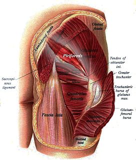
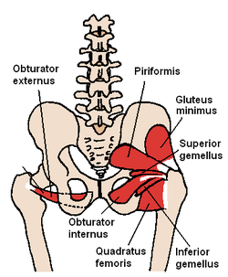
The piriformis muscle is a muscle in the gluteal region of the lower limbs. It is one of the six muscles in the lateral rotator group.

Coccygeus muscle Muscle of the lower back arising by its apex from the spine of the ischium

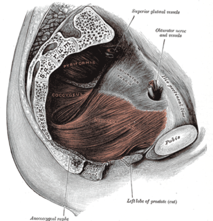
The coccygeus muscle or ischiococcygeus is a muscle of the pelvic floor, located posterior to levator ani and anterior to the sacrospinous ligament.

External obturator muscle One of six small hip muscles in the lateral rotator group

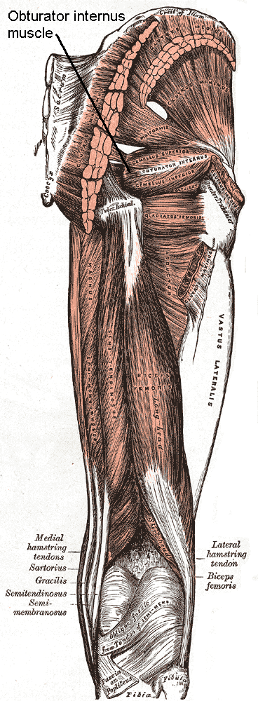
The external obturator muscle, obturator externus muscle is a flat, triangular muscle, which covers the outer surface of the anterior wall of the pelvis.

The semimembranosus muscle is the most medial of the three hamstring muscles in the thigh. It is so named because it has a flat tendon of origin. It lies posteromedially in the thigh, deep to the semitendinosus muscle. It extends the hip joint and flexes the knee joint.

Ischium

The ischium forms the lower and back part of the hip bone.

Pubis (bone)

In vertebrates, the pubic bone is the most forward-facing of the three main bones making up the pelvis. The left and right pubic bones are each made up of three sections, a superior ramus, inferior ramus, and a body.

Superior gluteal artery

The superior gluteal artery is the largest and final branch of the internal iliac artery. It is the continuation of the posterior division of that vessel. It is a short artery which runs backward between the lumbosacral trunk and the first sacral nerve. It divides into a superficial and a deep branch after passing out of the pelvis above the upper border of the piriformis muscle.

Obturator artery

The obturator artery is a branch of the internal iliac artery that passes antero-inferiorly on the lateral wall of the pelvis, to the upper part of the obturator foramen, and, escaping from the pelvic cavity through the obturator canal, it divides into both an anterior and a posterior branch.

Lesser sciatic foramen

The lesser sciatic foramen is an opening (foramen) between the pelvis and the back of the thigh. The foramen is formed by the sacrotuberous ligament which runs between the sacrum and the ischial tuberosity and the sacrospinous ligament which runs between the sacrum and the ischial spine.

Pudendal canal

The pudendal canal is an anatomical structure in the pelvis through which the internal pudendal artery, internal pudendal veins, and the pudendal nerve pass.

Obturator internus nerve

The nerve to obturator internus, also known as the obturator internus nerve, is a nerve that innervates the obturator internus and gemellus superior muscles.

Nerve to quadratus femoris

The nerve to quadratus femoris is a nerve that provides innervation to the quadratus femoris muscle and gemellus inferior muscle.

Lesser sciatic notch

Below the ischial spine is a small notch, the lesser sciatic notch; it is smooth, coated in the recent state with cartilage, the surface of which presents two or three ridges corresponding to the subdivisions of the tendon of the Obturator internus, which winds over it.

Pelvic fascia

The pelvic fasciae are the fascia of the pelvis and can be divided into:

Outline of human anatomy Overview of and topical guide to human anatomy

The following outline is provided as an overview of and topical guide to human anatomy:

Hip bone

The hip bone is a large irregular bone, constricted in the center and expanded above and below. In some vertebrates it is composed of three parts: the ilium, ischium, and the pubis.

References

This article incorporates text in the public domain from page 477 of the 20th edition of Gray's Anatomy (1918)

  1. 1 2 3 4 5 6 7 8 Chiva, Luis M.; Magrina, Javier (2018-01-01), Ramirez, Pedro T.; Frumovitz, Michael; Abu-Rustum, Nadeem R. (eds.), "Chapter 2 - Abdominal and Pelvic Anatomy", Principles of Gynecologic Oncology Surgery, Elsevier, pp. 3–49, doi:10.1016/b978-0-323-42878-1.00002-x, ISBN   978-0-323-42878-1 , retrieved 2021-03-04
  2. Jacob, S. (2008-01-01), Jacob, S. (ed.), "Chapter 4 - Abdomen", Human Anatomy, Churchill Livingstone, pp. 71–123, doi:10.1016/b978-0-443-10373-5.50007-5, ISBN   978-0-443-10373-5 , retrieved 2021-03-04
  3. Ahmadian, Amir; Abel, Naomi; Dakwar, Elias (2015-01-01), Tubbs, R. Shane; Rizk, Elias; Shoja, Mohammadali M.; Loukas, Marios (eds.), "Chapter 35 - Injuries to the Nerves of the Abdominopelvic Region", Nerves and Nerve Injuries, San Diego: Academic Press, pp. 545–555, ISBN   978-0-12-802653-3 , retrieved 2021-02-27
  4. de Groat, William C.; Yoshimura, Naoki (2015-01-01), Vodušek, David B.; Boller, François (eds.), "Chapter 5 - Anatomy and physiology of the lower urinary tract", Handbook of Clinical Neurology, Neurology of Sexual and Bladder Disorders, Elsevier, 130: 61–108, doi:10.1016/b978-0-444-63247-0.00005-5, PMID   26003239 , retrieved 2021-02-27
  5. Midha, Rajiv (2008-01-01), Kim, Daniel H.; Midha, Rajiv; Murovic, Judith A.; Spinner, Robert J. (eds.), "2 - Mechanisms and pathology of injury", Kline and Hudson's Nerve Injuries (Second Edition), Edinburgh: W.B. Saunders, pp. 23–42, doi:10.1016/b978-0-7216-9537-2.50010-4, ISBN   978-0-7216-9537-2 , retrieved 2021-03-04
  6. Bouche, P. (2013-01-01), Said, Gérard; Krarup, Christian (eds.), "Chapter 19 - Compression and entrapment neuropathies", Handbook of Clinical Neurology, Peripheral Nerve Disorders, Elsevier, 115, pp. 311–366, doi:10.1016/b978-0-444-52902-2.00019-9 , retrieved 2021-03-04
  7. Richenberg, Jonathan L. (2011-01-01), Allan, Paul L.; Baxter, Grant M.; Weston, Michael J. (eds.), "CHAPTER 29 - Ultrasound of the bladder", Clinical Ultrasound (Third Edition), Edinburgh: Churchill Livingstone, pp. 550–571, doi:10.1016/b978-0-7020-3131-1.00029-8, ISBN   978-0-7020-3131-1 , retrieved 2021-03-04