
The foot is an anatomical structure found in many vertebrates. It is the terminal portion of a limb which bears weight and allows locomotion. In many animals with feet, the foot is a separate organ at the terminal part of the leg made up of one or more segments or bones, generally including claws and/or nails.

The leg is the entire lower limb of the human body, including the foot, thigh or sometimes even the hip or buttock region. The major bones of the leg are the femur, tibia, and adjacent fibula. The thigh is between the hip and knee, while the calf (rear) and shin (front) are between the knee and foot.

In human anatomy, the fibularis longus is a superficial muscle in the lateral compartment of the leg. It acts to tilt the sole of the foot away from the midline of the body (eversion) and to extend the foot downward away from the body at the ankle.

The metatarsal bones or metatarsus are a group of five long bones in the midfoot, located between the tarsal bones and the phalanges (toes). Lacking individual names, the metatarsal bones are numbered from the medial side : the first, second, third, fourth, and fifth metatarsal. The metatarsals are analogous to the metacarpal bones of the hand. The lengths of the metatarsal bones in humans are, in descending order, second, third, fourth, fifth, and first. A bovine hind leg has two metatarsals.

The tibial nerve is a branch of the sciatic nerve. The tibial nerve passes through the popliteal fossa to pass below the arch of soleus.

In human anatomy, the dorsal interossei of the foot are four muscles situated between the metatarsal bones.

In human anatomy, the fibularis brevis is a muscle that lies underneath the fibularis longus within the lateral compartment of the leg. It acts to tilt the sole of the foot away from the midline of the body (eversion) and to extend the foot downward away from the body at the ankle.
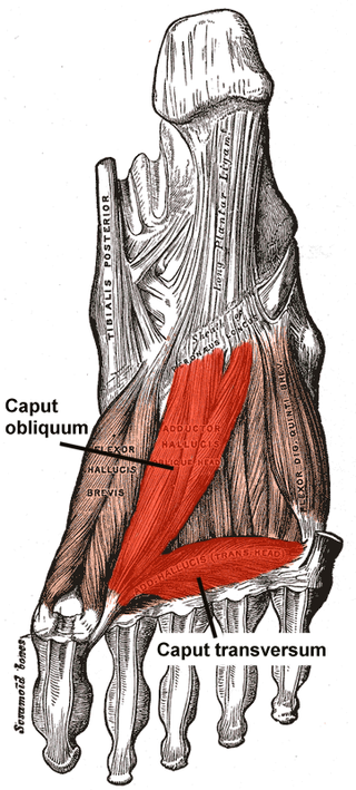
The Adductor hallucis arises by two heads—oblique and transverse and is responsible for adducting the big toe. It has two heads, both are innervated by the lateral plantar nerve.
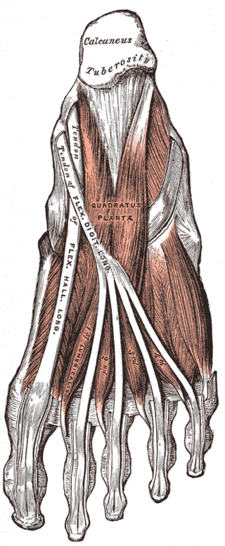
The quadratus plantae is separated from the muscles of the first layer by the lateral plantar vessels and nerve. It acts to aid in flexing the 2nd to 5th toes and is one of the few muscles in the foot with no homolog in the hand.
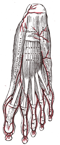
The abductor digiti minimi is a muscle which lies along the lateral (outer) border of the foot, and is in relation by its medial margin with the lateral plantar artery, vein and nerves.

The medial plantar nerve is the larger of the two terminal divisions of the tibial nerve, which accompanies the medial plantar artery.

The lateral plantar artery, much larger than the medial, passes obliquely lateralward and forward to the base of the fifth metatarsal bone.
The medial plantar artery, much smaller than the lateral plantar artery, passes forward along the medial side of the foot.
The deep plantar artery descends into the sole of the foot, between the two heads of the 1st interosseous dorsalis, and unites with the termination of the lateral plantar artery, to complete the plantar arch.
The lateral tarsal artery arises from the dorsalis pedis, as that vessel crosses the navicular bone; it passes in an arched direction lateralward, lying upon the tarsal bones, and covered by extensor hallucis brevis and extensor digitorum brevis; it supplies these muscles and the articulations of the tarsus, and receives the arcuate over the base of the fifth metatarsal. It may receive contributions from branches of the anterior lateral malleolar and the perforating branch of the peroneal artery directed towards the joint capsule, and from the lateral plantar arteries through perforating arteries of the foot.
The arcuate artery of the foot arises from dorsalis pedis slightly anterior to the lateral tarsal artery, specifically over the naviculocuneiform joint; it passes lateralward, over the bases of the lateral four metatarsal bones, beneath the tendons of the extensor digitorum brevis, its direction being influenced by its point of origin; and it terminates in the lateral tarsal artery. It communicates with the plantar arteries through the perforating arteries of the foot.
The plantar arch is a circulatory anastomosis formed from:

The second metatarsal bone is a long bone in the foot. It is the longest of the metatarsal bones, being prolonged backward and held firmly into the recess formed by the three cuneiform bones. The second metatarsal forms joints with the second proximal phalanx through the metatarsophalangeal joint, the cuneiform bones, third metatarsal and occasionally the first metatarsal bone.
The arcuate artery of the foot gives off the second, third, and fourth dorsal metatarsal arteries, which run forward upon the corresponding Interossei dorsales; in the clefts between the toes, each divides into two dorsal digital branches for the adjoining toes.

The proper plantar digital nerves of medial plantar nerve are nerves of the foot. They primarily arise from the medial plantar nerve's superficial and deep branches. The superficial branch of the medial plantar nerve turns into a proper digital nerve and is responsible for supplies the medial side of the great toe.
