Tail-shaped objects are sometimes referred to as "caudate" (e.g. caudate lobe, caudate nucleus), and the body part associated with or proximal to the tail are given the adjective "caudal" (which is considered a more precise anatomical terminology).
Function
Vulpes lagopus (Arctic fox) sleeping with its tail wrapped as a blanket.
Many animals use their tail for utility purposes, for example many grazing animals, such as horses and oxen, use their tails to drive away parasiticflies and sweep off other biting insects.[6][7] Some animals with broad, furry tails (e.g. foxes) often wrap the tail around the body as means of thermal insulation like a blanket.
Tails are also used for communication and signalling. Most canines use their tails to communicate mood and intention.[9] Some deer species flash the white underside of their tails to warn other nearby deer of possible danger,[10] beavers slap the water with their tails to indicate danger,[11]felids raise and quiver their tails while scent-marking,[12] and canids (including domestic dogs) indicate emotions through the positioning and movement of their tails.[13]Rattlesnakes perform tail vibration to generate a distinct rattling noise that signals aggression and warns potential predators to stay away.
Some species of lizard (e.g. geckos) can self-amputate ("cast") their tails from their bodies to help them escape predators, which are either distracted by the wriggling detached tail or only manages to seize the severed tail while the lizard flees. Tails cast in this manner generally grow back over time, though the replacement is typically darker in colour than the original and contains only cartilage, not bone.[14] Various species of rat demonstrate a similar function with their tails, known as degloving, in which the outer layer is shed in order for the animal to escape from a predator.[15]
Most birds' tails end in long feathers called rectrices. These feathers are used as a rudder, helping the bird steer and maneuver in flight; they also help the bird to balance while it is perched.[16] In some species—such as birds of paradise, lyrebirds, and most notably peafowl—modified tail feathers play an important role in courtship displays.[17] The extra-stiff tail feathers of other species, including woodpeckers and woodcreepers, allow them to brace themselves firmly against tree trunks.[18]
Human tails
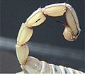
Tail-like structure on a female newborn from coccyx protrusion
In humans, tail bud refers to the part of the embryo which develops into the end of the spine.[19] However, this is not a tail.[20] Infrequently, a child is born with a "soft tail", which contains no vertebrae, but only blood vessels, muscles, and nerves, but this is regarded as an abnormality rather than a vestigial true tail, even when such an appendage is located where the tail would be expected.[21][22] Fewer than 40 cases have been reported of infants with "true tails" containing the caudal vertebrae, a result of atavism.[23]
In 2024, scientists claimed to have found a genetic mutation that contributed to the loss of the tail in the common ancestor of humans and other apes.[24][25]
Humans have a "tailbone" (the coccyx) attached to the pelvis; it comprises fused vertebrae, usually four, at the bottom of the vertebral column. It does not normally protrude externally – humans are an acaudal (or acaudate) species (i.e., tailless).
↑Mackenzie, SJ (2015). "Innervation and function of rat tail muscles for modeling cauda equina injury and repair". Muscle and Nerve. 52 (1): 94–102. doi:10.1002/mus.24498. PMID25346299. S2CID40356618.
↑"Tail Bud". Merriam Webster. Retrieved 4 June 2020.
↑"Developmental Stages in Human Embryos: Stage 16". the Endowment for Human Development. Retrieved 4 June 2020. What Kunitomo (1918) designated the "longest tail" at stage 16 is nothing of the kind but is merely the caudal end of the embryo, which will develop into the coccygeal region.
This page is based on this Wikipedia article Text is available under the CC BY-SA 4.0 license; additional terms may apply. Images, videos and audio are available under their respective licenses.